AntiNMDA
35.7K views | +1 today
Follow
 
Scooped by Nesrin Shaheen
onto AntiNMDA
Scoop.it!

Imaging Review of Paraneoplastic Neurologic Syndromes | American Journal of Neuroradiology

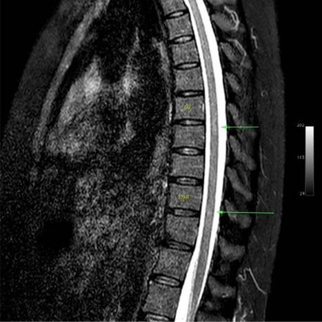
Abstract SUMMARY: Paraneoplastic syndromes are systemic reactions to neoplasms mediated by immunologic or hormonal mechanisms. The most well-recognized paraneoplastic neurologic syndrome, both clinically and on imaging, is limbic encephalitis. However, numerous additional clinically described syndromes affect the brain, spinal cord, and peripheral nerves. Many of these syndromes can have imaging findings that, though less well described, are important in making the correct diagnosis. Moreover, imaging in these syndromes frequently mimics more common pathology, which can be a diagnostic challenge for radiologists. Our goal is to review the imaging findings of paraneoplastic neurologic syndromes, including less well-known entities and atypical presentations of common entities. Specifically, we discuss limbic encephalitis, paraneoplastic cerebellar degeneration, paraneoplastic brain stem encephalitis, cranial neuropathy, myelitis, and polyneuropathy. We also demonstrate common diagnostic pitfalls that can be encountered when imaging these patients. ABBREVIATIONS: ANNA antineuronal nuclear autoantibody PNS paraneoplastic neurologic syndrome PCD paraneoplastic cerebellar degeneration Paraneoplastic syndromes (PNSs) result from systemic reactions to neoplasms, often mediated by immunologic or hormonal mechanisms. PNSs include limbic encephalitis, encephalomyelitis, paraneoplastic cerebellar degeneration (PCD), brain stem encephalitis, polyneuropathy, stiff-person syndrome, retinopathy, myasthenia gravis, Lambert-Eaton myasthenic syndrome, and enteric nervous system dysfunction (Fig 1). These syndromes are often associated with serum or CSF positivity of onconeuronal or neuronal cell surface antibodies. Onconeuronal antibodies are more directly associated with underlying neoplasms and cause neuronal dysfunction by recruitment of cytotoxic T cells. In contrast, neuronal cell surface antibodies are less commonly associated with an underlying neoplasm and mediate pathology by directly binding to neurons. PNSs can occur in the presence or absence of paraneoplastic antibodies and are ultimately a clinical diagnosis. Conversely, antibody-mediated neurologic syndromes can occur in the absence of malignancy, though these are separately classified.1 FIG 1. Illustration of the diverse array of paraneoplastic neurologic syndromes affecting a wide variety of anatomic structures. Most of these syndromes (in italics) can have salient imaging findings that can be important in making the correct diagnosis. Used with permission of Mayo Foundation for Medical Education and Research, all rights reserved. Many PNSs have salient imaging features. Although some of these are well described, particularly those of limbic encephalitis, others are not. Additionally, the spectrum of imaging findings for many PNSs is more variable than what is currently reported in the literature, which can frequently lead to incorrect or delayed diagnoses. Imaging is frequently obtained before the formal diagnosis of a PNS or underlying malignancy.2 Therefore, it is important to be aware of imaging features of these syndromes and common pitfalls. This review illustrates the typical imaging findings of paraneoplastic neurologic syndromes in a pictorial essay format and briefly discusses the differential diagnosis for each syndrome when encountered on imaging (On-line Table). Although prior reviews have comprehensively discussed the clinical features of these syndromes, their associated imaging features have not been widely demonstrated, with the notable exception of limbic encephalitis. The patients presented were ultimately diagnosed with paraneoplastic syndromes based on a combination of clinical, laboratory, and imaging findings. Several of the patients also highlight potential imaging pitfalls that can obfuscate the correct diagnosis. PARANEOPLASTIC NEUROLOGIC SYNDROMES BY ANATOMIC LOCATION Brain: Limbic Encephalitis Limbic encephalitis refers to inflammatory changes involving the limbic system, which includes the hippocampus, amygdala, hypothalamus, and cingulate cortex. Symptoms include mood and behavioral changes, cognitive dysfunction, memory loss, and seizure activity. Limbic encephalitis may be associated with various onconeuronal paraneoplastic antibodies, including antineuronal nuclear autoantibody type 1(ANNA-1)/anti-Hu, anti-collapsin response mediator protein-5 (CRMP5)/anti-CV2, and anti-Ma2. Although the syndromes associated with these antibodies all characteristically involve the limbic system, some can have extralimbic involvement. For example, anti-Ma2 can involve the brain stem and cerebellum, and anti-CRMP5 can have spinal cord involvement. Common tumor associations for limbic encephalitis in general include small cell lung cancer and breast cancer. Alternatively, limbic encephalitis can be seen in association with nonparaneoplastic neuronal cell surface antibodies, causing an autoimmune encephalitis. These include leucine-rich glioma-inactivated-1 (LGI1) autoantibodies, GAD65 autoantibodies, and anti–contactin-associated proteinlike 2 (CASPR2) antibodies.3,4 Patients with autoimmune encephalitis often have typical limbic system involvement, but sometimes different imaging findings such as subcortical T2 hyperintensities are seen.5 Imaging findings in paraneoplastic limbic encephalitis are ultimately not reliably distinguishable from nonparaneoplastic autoimmune causes, so it is important to be aware of autoantibodies that are more associated with nonparaneoplastic limbic encephalitis. Additionally, even autoimmune encephalitides associated with a typically nonparaneoplastic antibody have been uncommonly seen with underlying malignancy. Therefore, malignancy screening is nearly always appropriate.6 Typical imaging findings of limbic encephalitis include T2 hyperintensity and swelling of the mesial temporal lobes with FDG avidity on PET (Fig 2A, -B), sometimes with associated enhancement. These findings can also involve other parts of the limbic system. For example, paraneoplastic limbic encephalitis can have striking involvement of the hypothalamus and mammillary bodies with relative sparing of the remaining limbic system (Fig 2C–F). Sometimes limbic encephalitis can have a masslike appearance that can be mistaken for tumors such as low- or high-grade gliomas or even lymphoma (Fig 3A–C). The imaging findings in such cases can dramatically improve with treatment, and FDG hypometabolism may be seen after recovery (Fig 3D–F). Overall, limbic encephalitis has more complex imaging features than previously appreciated and should be considered even in cases that do not involve typical areas such as the hippocampus and amygdala. Atypical patterns of involvement can be seen in nonparaneoplastic limbic encephalitis as well.1,6 FIG 2. Classic limbic encephalitis (A and B). A 75-year-old healthy man presented with abrupt onset of memory loss and staring spells over several days. CSF analysis was positive for LGI1 autoantibodies. FDG-PET/CT axial image (A) shows marked hypermetabolism in the bilateral mesial temporal lobes (A, arrows). Axial double inversion recovery MR imaging image (B) shows corresponding increased T2 signal in both mesial temporal lobes (B, arrows). Atypical limbic encephalitis (C–F). A 29-year-old man with a history of resected nongerm cell testicular cancer presented months after treatment with worsening memory, diminished executive function, hypogonadism, and hypothyroidism. CSF analysis showed increased total protein but was otherwise normal. Axial FLAIR (C) and coronal T1-weighted postcontrast (D) images show abnormal T2 signal in the hypothalamus (C, arrows) and enhancement of the mammillary bodies (D, arrows). He was diagnosed with paraneoplastic limbic encephalitis with atypical involvement of the hypothalamus and mammillary bodies. He opted for observation with symptomatic improvement over months. Follow-up MR imaging shows improved mild residual hypothalamic T2 signal (E, arrows) and decreased enhancement of the mammillary bodies (F, arrows). FIG 3. Limbic encephalitis mimicking a tumor. A 31-year-old man presented after being found unconscious while doing repair work on his home. He had multiple neurologic symptoms, including impaired thermoregulation, hypersexuality, and depression. Axial FLAIR (A) and coronal T1-weighted postgadolinium (B) images show extensive increased T2 signal and masslike enhancement centered in the region of the hypothalamus (A and B, arrows). The findings were initially considered worrisome for a neoplastic process, such as lymphoma, versus an inflammatory lesion, such as lymphocytic hypophysitis. Biopsy of this region showed a nonspecific macrophage-rich demyelinating lesion with no evidence of tumor. He was later found to have anti-Ma2 serum positivity and a mediastinal germ cell tumor. Images of the brain from his staging FDG-PET/CT show asymmetric FDG uptake in the left mesial temporal lobe (C, arrows), separate from the affected regions on his MR imaging. He was treated with chemotherapy and high-dose corticosteroids with gradual improvement in his symptoms. Axial FLAIR (D) and coronal postgadolinium (E) MR imaging obtained 1 year later show marked improvement in the prior findings, with mild residual hypothalamic T2 signal (D, arrows) and mild enhancement of the tuber cinereum (E, arrows). His subsequent FDG-PET/CT shows decreased FDG avidity in the previously involved left mesial temporal lobe (F, arrows). Ultimately, his intracranial findings were consistent with limbic encephalitis rather than malignancy. The primary differential to exclude in limbic encephalitis with mesial temporal lobe involvement is herpes simplex viral encephalitis. Ancillary findings such as cortical petechial hemorrhages, or rarely intracerebral hematomas, can suggest herpes infection. CSF testing and prophylactic antiviral treatment are necessary if infection is a clinical possibility. Other differential considerations include status epilepticus, neurosyphilis, and low- or high-grade glioma.4 When limbic encephalitis has hypothalamic involvement, inflammatory conditions such as sarcoidosis or lymphocytic hypophysitis should be considered. Diagnostic criteria for limbic encephalitis include subacute symptom onset over less than 3 months, limbic system abnormalities on brain MR imaging, EEG showing epileptic or slow-wave activity in the temporal lobes or CSF pleocytosis, and reasonable exclusion of other etiologies.7 Thus, the work-up for limbic encephalitis, in addition to a thorough neurologic history and physical examination, typically includes brain MR imaging, paraneoplastic antibody testing, EEG, and oncologic work-up, including FDG-PET/CT. Brain biopsy is rarely needed but can be difficult to avoid when imaging shows a masslike abnormality. After the diagnosis is established, the most important next step is management of any underlying malignancy.8 However, immunosuppression and plasma exchange can be helpful, too. Brain: Paraneoplastic Cerebellar Degeneration PCD is clinically characterized by limb and truncal ataxia, nystagmus, and dysarthria.9 These symptoms are often progressive over weeks to months. Pathologically, patients have progressive loss of Purkinje cells in the cerebellum.10 CSF analysis can show pleocytosis and elevated oligoclonal bands. PCD is associated with a variety of paraneoplastic antibodies, including anti-Yo (Purkinje cell autoantibody type-1 [PCA-1]), anti-Ri (ANNA-2), and anti-Purkinje cell autoantibody-Tr/DNER (delta/notchlike epidermal growth factor-related receptor). About 60% of cases have an associated onconeuronal antibody.9 Common associated malignancies include ovarian cancer, breast cancer, and Hodgkin lymphoma. Imaging findings of PCD depend on the phase of the syndrome. In the acute phase, increased T2 signal within the cerebellar hemispheres can be seen (Fig 4A, -B). At this stage, the differential diagnosis includes infectious or inflammatory cerebellitis. In the chronic phase, T2 hyperintensity improves, and cerebellar FDG-PET hypometabolism, atrophy, or both may be seen (Fig 4C–F). The differential diagnosis for chronic PCD includes alcoholic degeneration, neurodegenerative disorders such as multiple system atrophy (cerebellar subtype), and sequelae from infectious/inflammatory cerebellitis. Careful attention to patient history, such as prior alcohol use disorder or cerebellar infection, is most important in distinguishing these entities. FIG 4. Subacute paraneoplastic cerebellar ataxia (A and B). A 58-year-old woman presented with headaches, vertigo, and ataxia that had progressed over a period of weeks. CSF analysis showed a high titer of PCA-1 autoantibodies. After additional work-up, she was found to have metastatic breast adenocarcinoma. Axial FLAIR (A) and T2-weighted (B) images of the brain show marked T2 hyperintensity diffusely involving both cerebellar hemispheres (A and B, arrows). No enhancement was seen on postgadolinium images (not shown). The patient was diagnosed with paraneoplastic cerebellar degeneration. She had mild improvement in her neurologic symptoms with high-dose corticosteroids but remained wheelchair dependent. Chronic paraneoplastic cerebellar ataxia (C–F). A 56-year-old woman presented with progressive ataxia over a period of several months. Paraneoplastic serum antibody panel revealed anti-G-AchR (ganglionic acetylcholine receptor) antibodies, and CSF analysis revealed increased protein concentration and high IgG levels. Initial axial T2-weighted image of the brain (C) and subsequent axial T2-weighted image from an examination several months later (D) show interval development of mild left cerebellar atrophy (D, arrows). FDG-PET axial source image of the brain (E) and processed surface rendering of the brain compared with age-matched control participants (F) show marked hypometabolism within the left cerebellar hemisphere (E and F, arrows). No cerebral lesion was present to suggest crossed cerebellar diaschisis. Review of prior records noted an FDG-avid thyroid lesion found to be a follicular neoplasm on resection. She was diagnosed with paraneoplastic cerebellar degeneration with chronic atrophic features. Her symptoms have been stable on cyclophosphamide with pulsed corticosteroids. As with limbic encephalitis, brain MR imaging, paraneoplastic antibody testing, and oncologic work-up are key to diagnosis. Pelvic sonography or MR imaging should also be considered in women because PCD is associated with gynecologic malignancies.11 Treatment of any identifiable malignancy is critical, but immunosuppressive agents are also used, ranging from corticosteroids to cyclophosphamide for severe cases. Treatment response is generally poor if antibodies to intracellular antigens are detected (eg, PCA-1/anti-Yo), and most of these patients progress to wheelchair dependence. If an antibody to a cell surface antigen is instead found (eg, metabotropic glutamate receptor [mGluR1]), the prognosis is better. Brain: Brain Stem Encephalitis Brain stem encephalitis, or rhombencephalitis, refers to inflammation predominantly involving the brain stem, though the cerebellar peduncles and hemispheres can be affected too. Paraneoplastic brain stem encephalitis has a variable clinical presentation that may include ataxia, dysarthria, and ophthalmoplegia.12 It is most commonly associated with the anti-Ma2 antibody but has also recently been identified with antibodies to Kelchlike protein 11.13 The syndrome is most associated with testicular germ cell tumors such as seminomas.9 However, it can also be seen with neuroendocrine and other tumors.14 Imaging findings can be normal in these patients. When present, findings vary depending on the phase of illness. Acutely, T2 hyperintensity and/or enhancement of the cerebellum, cerebellar peduncles, and brain stem are seen. Importantly, there can be isolated involvement of the brain stem in the acute phase, which can be mistaken for other entities, including tumor (Fig 5A, -B). As the disease progresses, cerebellar and brain stem atrophy are often seen. Additionally, cruciform T2 hyperintensity in the pons and T2 hyperintensity in the middle cerebellar peduncles can occur with this syndrome secondary to Wallerian degeneration, quite similar to the imaging findings seen in multiple system atrophy (Fig 5C, -D). Hypertrophic olivary degeneration can also be seen as a result of paraneoplastic brain stem encephalitis (Fig 5E, -F), presumably caused by disruption of the dentato-rubro-olivary pathway. FIG 5. Paraneoplastic brain stem encephalitis mimicking a tumor (A–D). A 29-year-old man presented with tinnitus, hearing loss, diplopia, and imbalance that had progressed over at least 1 year. After extensive clinical work-up, the patient was diagnosed with metastatic seminoma to the mediastinum and noted to have elevated CSF IgG. Serum and CSF were positive for Kelchlike protein 11 autoantibodies. Sagittal T1-weighted postcontrast (A) and sagittal FLAIR (B) images show enhancing, T2 hyperintense lesions in the midbrain and tectum (A and B, arrows), initially believed to be worrisome for metastases. He was treated with chemotherapy, and his symptoms transiently improved but then worsened over the next several years. Sagittal T1-weighted postcontrast (C) and axial FLAIR (D) images from a subsequent MR imaging 4 years later show new marked cerebellar and brain stem atrophy (C, arrows), as well as new T2 signal in the middle cerebellar peduncles (D, arrows) and cruciform T2 signal in the pons (D, arrowhead). He was diagnosed with paraneoplastic brain stem encephalitis rather than multiple system atrophy given the clinical context of the findings. The patient’s symptoms mildly improved after treatment with multiple immunosuppressive agents. Brain stem encephalitis associated with hypertrophic olivary degeneration (E and F). A 45-year-old man presented with progressive aural fullness and hearing loss, initially thought to be caused by otitis media. Over the next 2 years, he had progressive gait ataxia and diplopia. CSF analysis showed antibodies to Kelchlike protein 11. Testicular sonography, performed as part of his oncologic work-up, showed a right testicular mass. Right orchiectomy revealed a germ cell neoplasm. Axial FLAIR images (E and F) from his brain MR imaging show enlargement and T2 hyperintensity of both inferior olives (E, arrows) consistent with bilateral hypertrophic olivary degeneration. Additionally, there is patchy T2 hyperintensity in the cerebellar vermis (F, arrows). He was diagnosed with paraneoplastic rhombencephalitis with associated hypertrophic olivary degeneration. He has had a partial response to multiple immunotherapies. Differential considerations for the acute phase are broad, though certain conditions are known to have a predilection for the brain stem. These include Listeria and herpes simplex virus infection as well as inflammatory disorders such as Behçet disease, systemic lupus erythematosus, Bickerstaff encephalitis, or Miller Fischer syndrome.12,15 Demyelinating diseases such as multiple sclerosis, neuromyelitis optica spectrum disorder, and myelin oligodendrocyte glycoprotein antibody–associated disease are additional considerations. In the chronic phase, this syndrome can be mistaken for multiple system atrophy and other neurodegenerative disorders. In addition to brain MR imaging and usual paraneoplastic antibody screening, screening for the newly discovered KLHL11 autoantibodies should be done when diagnosing these patients. Furthermore, testicular sonography should be done in men given the association with testicular seminoma.11 The mainstay of management is treatment of any identifiable malignancy, with immunomodulatory therapy used in refractory cases. Head: Cranial Neuropathy Paraneoplastic cranial neuropathies are rare but have been previously described in several reports.16,17 Symptoms depend on the cranial nerves involved. For example, deteriorating balance and sensorineural hearing loss have been observed with paraneoplastic involvement of the eighth cranial nerves.17 Symptoms may be more extensive when there is involvement of multiple cranial nerves, as seen in 1 report of paraneoplastic neuropathy of the third, sixth, and seventh cranial nerves in a patient with gallbladder carcinoma.18 Anti-Hu/ANNA-1 and the newly described antibodies to Kelchlike protein-11 associated with seminoma are especially associated with paraneoplastic cranial neuropathy. Possible imaging findings include enhancement and/or enlargement of the affected cranial nerves (Fig 6). The differential diagnosis includes infection, acute inflammatory demyelinating polyneuropathy (Guillain-Barré syndrome), and chronic inflammatory demyelinating polyneuropathy.19,20 Entities such as neurosarcoidosis, lymphoma, and carcinomatous meningitis tend to have more nodular enhancement and likely other areas of involvement, whereas chronic inflammatory demyelinating polyneuropathy tends to have mainly nerve root enlargement without enhancement. Notably, leptomeningeal metastases are a frequent clinical concern in patients with cancer who have new cranial neuropathy, so imaging can play an important role in potentially suggesting a paraneoplastic rather than malignant cause (Fig 6). FIG 6. Paraneoplastic cranial neuropathy mimicking leptomeningeal carcinomatosis. A 54-year-old woman recently diagnosed with small cell lung cancer presented with diplopia that slowly progressed over a period of days. Her examination showed left eye ptosis and gaze abnormalities consistent with oculomotor palsy. The primary clinical concern was metastatic leptomeningeal carcinomatosis. Lumbar puncture showed elevated CSF IgG index and IgG synthesis rate with no specific findings for malignancy. No specific paraneoplastic antibodies were detected. Axial T1-weighted pre- (A) and postcontrast (B) images and the coronal postcontrast image (C) from her brain MR imaging show enlargement and enhancement of the left third cranial nerve cisternal segment (B and C, arrows) and mild enhancement of the right third cranial nerve cisternal segment (C, arrowhead). Coronal T1-weighted image (D) confirms mild enlargement of the left third cranial nerve (arrow). Based on the lack of other widespread intracranial disease, non-nodular nerve enhancement, and CSF findings, she was diagnosed with paraneoplastic cranial neuropathy and had rapid symptomatic improvement with methylprednisolone. Diagnosis requires a thorough neurologic work-up, and ophthalmologic consultation is helpful if ophthalmoplegia is identified. Brain MR imaging with thin-section imaging is particularly useful to assess the cranial nerves and patterns of pathologic enhancement. The treatment for paraneoplastic cranial neuropathy is not well studied and mainly involves management of the underlying malignancy. In our experience, patients have improved with short-term corticosteroids. Spine: Myelitis Paraneoplastic myelitis is a rare but likely underreported entity. It may occur in isolation or in conjunction with other involved regions (eg, paraneoplastic encephalomyelitis).9,21 Even in such cases, it differs from limbic encephalitis insofar as it has more generalized cerebral abnormalities not confined to the limbic system. Patients can have an acute myelitis or more insidious progressive myelopathy with combinations of weakness, numbness, and bowel or bladder dysfunction. Other symptoms can include headache, seizures, and agitation, attributable to the associated intracranial involvement.6 A variety of paraneoplastic antibodies can be seen, but anti-CRMP5/anti-CV2 and anti-amphiphysin are the most common. Small cell lung and breast cancer are the most frequently associated malignancies. A paraneoplastic aquaporin-4 IgG seropositive neuromyelitis optica spectrum disorder is also recognized.22 When confined to the spinal cord, imaging findings include longitudinally extensive T2 hyperintensity and enhancement (Fig 7). Tract-specific abnormalities, particularly along the lateral columns, are characteristic (Fig 7C, -G). Abnormal FDG uptake can also be present in the involved area of the spinal cord with this syndrome (Fig 7E), which could potentially be mistaken for metastatic tumor involvement.23 Differential considerations for this entity include neuromyelitis optica spectrum disorder, neurosarcoidosis, spinal dural AVF, radiation myelitis, and cord infarct. The presence of perimedullary flow voids usually suggests an AVF, whereas preferential involvement of the anterior horns often suggests cord infarct. Multiple sclerosis would not generally have longitudinally extensive involvement. FIG 7. Paraneoplastic myelitis (A–E). A 52-year-old woman recently diagnosed with small cell lung cancer presented with progressive bilateral lower extremity weakness over weeks. Lumbar puncture was remarkable for elevated CSF IgG index and IgG synthesis rate, though no specific paraneoplastic antibody was identified. Sagittal T1-weighted pre- (A) and postcontrast (B) images and an axial postcontrast image (C) from her thoracic spine MR imaging show a long segment of enhancement involving the lateral columns of the thoracic spinal cord. Corresponding axial T2-weighted (D) and FDG-PET/CT (E) images show increased T2 signal (D, arrows) and marked abnormal FDG uptake (E, arrows) in the spinal cord. She was diagnosed with paraneoplastic myelopathy and had transient improvement with methylprednisolone. Paraneoplastic myelitis with treatment response (F–I). A 64-year-old woman recently diagnosed with small cell lung cancer presented with gait difficulties and progressive bilateral lower extremity weakness lasting several months. Serum and CSF were positive for CRMP5 autoantibodies. Sagittal T2-weighted (F) and axial T1-weighted postcontrast (G) images of the cervical cord show increased T2 signal (F, arrows) and enhancement of the lateral columns (G, arrows). She was treated with 6 months of cyclophosphamide and had partial symptomatic improvement. Follow-up sagittal T2-weighted image (H) shows near resolution of abnormal signal. There is mild residual patchy cord enhancement, greatest in the lateral columns (I, arrows). In addition to the usual paraneoplastic work-up, MR imaging of the entire spine is particularly helpful in these patients because of the longitudinally extensive nature of spinal cord involvement. Imaging of the brain is also usually indicated to assess for associated encephalitis, which is frequently seen with anti-CRMP5 syndromes. Typical treatment includes management of the underlying malignancy and immunosuppression. Despite treatment, the outcome is overall poor with most patients becoming wheelchair dependent. Although follow-up of patients with paraneoplastic myelitis is not well documented in the literature, clinical improvement and improvement in imaging findings can occur (Fig 7F–I). Spine: Polyneuropathy Paraneoplastic polyneuropathy is rare but usually manifests clinically as a sensory neuropathy.9 This is characterized by pain, paresthesias, and loss of vibratory sensation, often with ANNA-1/anti-Hu or anti-CRMP5 antibody positivity. Much less commonly, paraneoplastic syndromes can also have other combinations of motor, sensory, and autonomic deficits.24 As examples, paraneoplastic syndromes associated with Hodgkin lymphoma and prostate cancer causing motor and sensory deficits were recently reported.25-27 Autonomic dysfunction was also previously reported from a syndrome related to a carcinoid tumor.28 Reports of imaging findings of paraneoplastic polyneuropathy involvement are scarce. To our knowledge, there are only 5 published cases of paraneoplastic polyneuropathy showing cauda equina abnormalities. These have demonstrated variable patterns of enhancement, with 2 patients having ANNA-1 positivity.27,29-32 We have found that enhancement can predominantly involve the dorsal (Fig 8) or ventral nerve roots (Fig 9), and the pattern of enhancement does not always correlate with symptoms. The clinical presentation can frequently mimic Guillain-Barré syndrome with a similar rapidly ascending weakness (Fig 8). Additionally, polyneuropathy may be seen with osteosclerotic multiple myeloma in the setting of POEMS syndrome (polyneuropathy, organomegaly, endocrinopathy, monoclonal plasma proliferative disorder, skin changes).33 Although POEMS syndrome is not always described among the classic forms of PNS, many authors consider its peripheral nerve involvement to be paraneoplastic. The presence of sclerotic bone lesions and peripheral nerve root enhancement are the most important imaging features to suggest POEMS syndrome (Fig 10). FIG 8. Paraneoplastic polyneuropathy mimicking Guillain-Barré syndrome. A 69-year-old woman presented with rapidly progressive ascending weakness and paresthesias. CSF analysis was remarkable for albuminocytologic dissociation, and the patient was initially diagnosed with Guillain-Barré syndrome. Sagittal T1-weighted pre- (A) and postcontrast (B) and axial T1-weighted pre- (C) and postcontrast (D) images from the patient’s lumbar spine MR imaging show enhancement of the predominantly dorsal cauda equina nerve roots (B–D, arrowheads). There is relative sparing of the ventral nerve roots (D, arrows), somewhat atypical for Guillain-Barré syndrome. The patient’s CSF was later found to be ANNA-1/anti-Hu antibody positive, and she was ultimately found to have small cell lung cancer. FIG 9. Ventral predominant paraneoplastic polyneuropathy. A 29-year-old man with a history of mediastinal seminoma presented with progressive lower extremity weakness and frequent falls over months. Lumbar puncture showed elevated CSF IgG. Serum and CSF were positive for Kelchlike protein 11 autoantibodies. Sagittal T1-weighted pre- (A) and postcontrast (B) images, as well as corresponding axial T1-weighted pre- (C) and postcontrast (D) images from his lumbar spine MR imaging demonstrate smooth enhancement of the cauda equina nerve roots with a predilection for the ventral roots (B and D, arrows). There is clear sparing of the dorsal roots (B and D, arrowheads). He was diagnosed with paraneoplastic polyneuropathy and had mild symptomatic improvement with numerous immunosuppressive agents. FIG 10. POEMS syndrome. A 50-year-old woman presented with weakness, burning, and tingling in her extremities that had waxed and waned over 4 years. Axial T1-weighted pre- (A) and postcontrast (B) images from her lumbar spine MR imaging show diffuse enlargement and enhancement of the cauda equina nerve roots (B, arrowheads). Axial CT (C) and fused FDG-PET/CT (D) images show a non–FDG-avid lumbar vertebral sclerotic lesion (C and D, arrowheads) and an adjacent mildly FDG-avid sclerotic lesion (C and D, arrows). After additional work-up, she was diagnosed with osteosclerotic multiple myeloma, which is characterized by osteosclerotic lesions with variable FDG avidity. Her MR imaging findings were ultimately attributed to polyneuropathy in the setting of POEMS syndrome. Differential diagnoses for paraneoplastic polyneuropathy mainly include Guillain-Barré syndrome and infection. Guillain-Barré syndrome classically, but not always, has ventral predominant nerve enhancement. Chronic inflammatory demyelinating polyneuropathy could be considered but is less likely to show any enhancement and would have more prominent nerve enlargement. Entities such as neurosarcoidosis or leptomeningeal carcinomatosis tend to have more nodular enhancement. Ventral cauda equina enhancement has also recently been described in association with cord infarct.34 The diagnostic work-up should include a thorough history to elicit any antecedent infection that may suggest Guillain-Barré syndrome, a complete neurologic examination, paraneoplastic antibody testing, electromyography, and lumbar spine MR imaging. Treatment is not well studied, but we have observed symptomatic improvement with corticosteroids and plasma exchange. CONCLUSIONS Paraneoplastic neurologic syndromes can cause a wide range of imaging abnormalities throughout the central and peripheral nervous systems. Prior reports have focused on the imaging findings of limbic encephalitis, but as we have shown, PNSs involving other parts of the nervous system can have characteristic imaging findings. We have described the imaging findings of paraneoplastic limbic encephalitis, cerebellar degeneration, brain stem encephalitis, cranial neuropathy, myelitis, and polyneuropathy. Additionally, we have highlighted the fact that these entities can have variable imaging findings resulting in many potential diagnostic pitfalls. Because imaging features of each entity are sometimes nonspecific, consideration of paraneoplastic syndromes based on imaging findings combined with review of patient history are important. Although serum and CSF paraneoplastic antibody positivity can be helpful, they may be absent, as illustrated in many of our cases. Importantly, many of these syndromes respond to treatment of underlying malignancy or immunotherapy. Therefore, timely diagnosis is likely to significantly impact patient outcomes. Last, it should be noted that PNSs are relatively rare disorders compared with various conditions in the differential diagnosis. The diagnosis of PNS often requires exclusion of these more common entities. ACKNOWLEDGMENT The authors acknowledge the assistance of Desiree J. Lanzino, PT, PhD, in editing the manuscript. Footnotes Disclosures: Eoin Flanagan—UNRELATED: Other: Viela Bio, Comments: I am a site principal investigator in a placebo-controlled randomized controlled phase 3 trial on inebilizumab in neuromyelitis optica spectrum disorder. Indicates open access to non-subscribers at www.ajnr.org References 1.↵Kelley BP, Patel SC, Marin HL, et al. Autoimmune encephalitis: pathophysiology and imaging review of an overlooked diagnosis. AJNR Am J Neuroradiol 2017;38:1070–78 doi:10.3174/ajnr.A5086 pmid:28183838Abstract/FREE Full Text 2.↵Leypoldt F, Wandinger KP. Paraneoplastic neurological syndromes. Clin Exp Immunol 2014;175:336–48 doi:10.1111/cei.12185 pmid:23937626CrossRefPubMed 3.↵Lancaster E, Huijbers MG, Bar V, et al. Investigations of caspr2, an autoantigen of encephalitis and neuromyotonia. Ann Neurol 2011;69:303–11 doi:10.1002/ana.22297 pmid:21387375CrossRefPubMed 4.↵da Rocha AJ, Nunes RH, Maia AC Jr., et al. Recognizing autoimmune-mediated encephalitis in the differential diagnosis of limbic disorders. AJNR Am J Neuroradiol 2015;36:2196–205 doi:10.3174/ajnr.A4408 pmid:26381566Abstract/FREE Full Text 5.↵Fredriksen JR, Carr CM, Koeller KK, et al. MRI findings in glutamic acid decarboxylase associated autoimmune epilepsy. Neuroradiology 2018;60:239–45 doi:10.1007/s00234-018-1976-6 pmid:29353399CrossRefPubMed 6.↵Guerin J, Watson RE, Carr CM, et al. Autoimmune epilepsy: findings on MRI and FDG-PET. Br J Radiology 2019;92:20170869 doi:10.1259/bjr.20170869 pmid:30235015CrossRefPubMed 7.↵Dutra LA, Abrantes F, Toso FF, et al. Autoimmune encephalitis: a review of diagnosis and treatment. Arq Neuropsiquiatr 2018;76:41–49 doi:10.1590/0004-282X20170176 pmid:29364393CrossRefPubMed 8.↵Lancaster E. The diagnosis and treatment of autoimmune encephalitis. J Clin Neurol 2016;12:1–13 doi:10.3988/jcn.2016.12.1.1 pmid:26754777CrossRefPubMed 9.↵Hoftberger R, Rosenfeld MR, Dalmau J. Update on neurological paraneoplastic syndromes. Curr Opin Oncol 2015;27:489–95 doi:10.1097/CCO.0000000000000222 pmid:26335665CrossRefPubMed 10.↵Jarius S, Wildemann B. “Medusa head ataxia”: the expanding spectrum of Purkinje cell antibodies in autoimmune cerebellar ataxia. Part 3: anti-Yo/CDR2, anti-Nb/AP3B2, PCA-2, anti-Tr/DNER, other antibodies, diagnostic pitfalls, summary and outlook. J Neuroinflammation 2015;12:168 doi:10.1186/s12974-015-0358-9 pmid:26377319CrossRefPubMed 11.↵Mahta A, Vijayvergia N, Bhavsar TM, et al. Diagnostic approach to a patient with paraneoplastic neurological syndrome. World J Oncol 2012;3:243–46 doi:10.4021/wjon571w pmid:29147315CrossRefPubMed 12.↵Campos LG, Trindade RA, Faistauer A, et al. Rhombencephalitis: pictorial essay. Radiology Bras 2016;49:329–36 doi:10.1590/0100-3984.2015.0189 pmid:27818548CrossRefPubMed 13.↵Mandel-Brehm C, Dubey D, Kryzer TJ, et al. Kelch-like protein 11 antibodies in seminoma-associated paraneoplastic encephalitis. N Engl J Med 2019;381:47–54 doi:10.1056/NEJMoa1816721 pmid:31269365CrossRefPubMed 14.↵Boch M, Rinke A, Rexin P, et al. Paraneoplastic brainstem encephalitis in a patient with exceptionally long course of a metastasized neuroendocrine rectum neoplasm. BMC Cancer 2014;14:691 doi:10.1186/1471-2407-14-691 pmid:25244967CrossRefPubMed 15.↵Jubelt B, Mihai C, Li TM, et al. Rhombencephalitis/brainstem encephalitis. Curr Neurol Neurosci Rep 2011;11:543–52 doi:10.1007/s11910-011-0228-5 pmid:21956758CrossRefPubMed 16.↵Cheung SSL, Lau GKK, Chan KH, et al. Optic neuritis as the initial clinical presentation of limbic encephalitis: a case report. J Med Case Rep 2018;12:357 doi:10.1186/s13256-018-1893-7 pmid:30509326CrossRefPubMed 17.↵Renna R, Plantone D, Batocchi AP. Teaching NeuroImages: a case of hearing loss in a paraneoplastic syndrome associated with anti-Hu antibody. Neurology 2012;79:e134 doi:10.1212/WNL.0b013e31826e262b pmid:23045521CrossRefPubMed 18.↵Kaido M, Yuasa Y, Yamamoto T, et al. A case of possible paraneoplastic neurological syndrome presenting as multiple cranial nerve palsies associated with gallbladder cancer. Rinsho Shinkeigaku 2016;56:617–21 doi:10.5692/clinicalneurol.cn-000906 pmid:27498818CrossRefPubMed 19.↵Nanda SK, Jayalakshmi S, Ruikar D, et al. Twelfth cranial nerve involvement in Guillain Barre syndrome. J Neurosci Rural Pract 2013;4:338–40 doi:10.4103/0976-3147.118804 pmid:24250180CrossRefPubMed 20.↵Inoue H, Tsuboi Y, Tsugawa J, et al. Hypertrophic cranial nerve roots in CIDP. Neurology 2004;63:1481 doi:10.1212/01.wnl.0000137033.77605.b2 pmid:15505169FREE Full Text 21.↵Flanagan EP, McKeon A, Lennon VA, et al. Paraneoplastic isolated myelopathy: clinical course and neuroimaging clues. Neurology 2011;76:2089–95 doi:10.1212/WNL.0b013e31821f468f pmid:21670438Abstract/FREE Full Text 22.↵Pittock SJ, Lennon VA, de Seze J, et al. Neuromyelitis optica and non organ-specific autoimmunity. Arch Neurol 2008;65:78–83 doi:10.1001/archneurol.2007.17 pmid:18195142CrossRefPubMed 23.↵Keegan BM, Flanagan EP. In reply—Sensitivity of [18F]-fluorodeoxyglucose-positron emission tomography in patients with active myelopathy. Mayo Clin Proc 2014;89:859 doi:10.1016/j.mayocp.2014.03.009 pmid:24943702CrossRefPubMed 24.↵Graus F, Santamaría J, Obach J, et al. Sensory neuropathy as remote effect of cancer. Neurology 1987;37:1266–67 doi:10.1212/wnl.37.7.1266-b pmid:3037442FREE Full Text 25.↵Al IO, Koc B, Bayram C, et al. Variant Guillain-Barré syndrome in a patient with Hodgkin lymphoma: AMSAN. Turk Pediatri Ars 2019;53:263–66 doi:10.5152/TurkPediatriArs.2018.4763CrossRef 26.Choi JK, Kim WJ, Jeon JY. Paraneoplastic sensorimotor polyneuropathy in prostatic adenocarcinoma: a case report. Medicine (Baltimore) 2018;97:e0030 doi:10.1097/MD.0000000000010030 pmid:29642138CrossRefPubMed 27.↵Nomiyama K, Uchino A, Yakushiji Y, et al. Diffuse cranial nerve and cauda equina lesions associated with breast cancer. Clin Imaging 2007;31:202–05 doi:10.1016/j.clinimag.2007.01.006 pmid:17449382CrossRefPubMed 28.↵Veilleux M, Bernier JP, Lamarche JB. Paraneoplastic encephalomyelitis and subacute dysautonomia due to an occult atypical carcinoid tumour of the lung. Can J Neurol Sci 1990;17:324–28 doi:10.1017/s0317167100030675 pmid:2207890CrossRefPubMed 29.↵Kumar N, Dyck PJ. Hypertrophy of the nerve roots of the cauda equina as a paraneoplastic manifestation of lymphoma. Arch Neurol 2005;62:1776–77 doi:10.1001/archneur.62.11.1776 pmid:16286555CrossRefPubMed 30.Flanagan EP, Sandroni P, Pittock SJ, et al. Paraneoplastic lower motor neuronopathy associated with Hodgkin lymphoma. Muscle Nerve 2012;46:823–27 doi:10.1002/mus.23464 pmid:23055320CrossRefPubMed 31.Shibata M, Uchida M, Tsukagoshi S, et al. Anti-Hu antibody-associated paraneoplastic neurological syndrome showing peripheral neuropathy and atypical multifocal brain lesions. Intern Med 2015;54:3057–60 doi:10.2169/internalmedicine.54.4867 pmid:26631892CrossRefPubMed 32.↵Madhavan AA, Guerin JB, Eckel LJ, et al. Dorsal cauda equina nerve root enhancement on magnetic resonance imaging due to ANNA-1-associated paraneoplastic polyneuropathy. Neuroradiol J 2020 Apr 15 [Epub ahead of print] doi:10.1177/1971400920919689 pmid:32290766CrossRefPubMed 33.↵Paranavitane S, Gooneratne L, Chang T. Polyneuropathy, organomegaly, endocrinopathy, monoclonal band, and skin (POEMS) changes syndrome presenting with a pseudosensory level: a case report. J Med Case Rep 2019;13:384 doi:10.1186/s13256-019-2309-z pmid:31878962CrossRefPubMed 34.↵Diehn FE, Hunt CH, Lehman VT, et al. Vertebral body infarct and ventral cauda equina enhancement: two confirmatory findings of acute spinal cord infarct. J Neuroimaging 2015;25:133–35 doi:10.1111/jon.12058 pmid:24118402CrossRefPubMed Received May 19, 2020. Accepted after revision June 29, 2020. © 2020 by American Journal of Neuroradiology
No comment yet.
AntiNMDA
Your new post is loading...
Scooped by Nesrin Shaheen
Scoop.it!

Josep Dalmau receives the “Scientific Breakthrough 2023” Award from the American Brain Foundation

The accolade recognises the commitment of this Clínic Barcelona-IDIBAPS researcher to deepening our understanding of autoimmune neurological diseases such...
No comment yet.
Scooped by Nesrin Shaheen
Scoop.it!

IDIBAPS creates three multidisciplinary research programs to encourage collaboration among its groups

They are the Translational cancer research program, the Synaptic autoimmunity in neurology, psychiatry and cognitive neuroscience program and the Lymphoid...
No comment yet.
Scooped by Nesrin Shaheen
Scoop.it!

ExTINGUISH: A Beacon of Hope for NMDAR Encephalitis

No comment yet.
Scooped by Nesrin Shaheen
Scoop.it!

MR Imaging Findings in a Large Population of Autoimmune Encephalitis | American Journal of Neuroradiology

MR Imaging Findings in a Large Population of Autoimmune Encephalitis | American Journal of Neuroradiology | AntiNMDA | Scoop.it
Research ArticleAdult Brain MR Imaging Findings in a Large Population of Autoimmune Encephalitis S. Gillon, M. Chan, J. Chen, E.L. Guterman, X. Wu, C.M. Glastonbury and Y. Li American Journal of Neuroradiology July 2023, 44 (7) 799-806; DOI: https://doi.org/10.3174/ajnr.A7907 ArticleFigures & DataInfo & MetricsReferences PDF This article requires a subscription to view the full text. If you have a subscription you may use the login form below to view the article. Access to this article can also be purchased. AbstractBACKGROUND AND PURPOSE: Autoimmune encephalitis is a rare condition in which autoantibodies attack neuronal tissue, causing neuropsychiatric disturbances. This study sought to evaluate MR imaging findings associated with subtypes and categories of autoimmune encephalitis.MATERIALS AND METHODS: Cases of autoimmune encephalitis with specific autoantibodies were identified from the medical record (2009–2019). Cases were excluded if no MR imaging of the brain was available, antibodies were associated with demyelinating disease, or >1 concurrent antibody was present. Demographics, CSF profile, antibody subtype and group (group 1 intracellular antigen or group 2 extracellular antigen), and MR imaging features at symptom onset were reviewed. Imaging and clinical features were compared across antibody groups using χ2 and Wilcoxon rank-sum tests.RESULTS: Eighty-five cases of autoimmune encephalitis constituting 16 distinct antibodies were reviewed. The most common antibodies were anti-N-methyl-D-aspartate (n = 41), anti-glutamic acid decarboxylase (n = 7), and anti-voltage-gated potassium channel (n = 6). Eighteen of 85 (21%) were group 1; and 67/85 (79%) were group 2. The median time between MR imaging and antibody diagnosis was 14 days (interquartile range, 4–26 days). MR imaging had normal findings in 33/85 (39%), and 20/33 (61%) patients with normal MRIs had anti-N-methyl-D-aspartate receptor antibodies. Signal abnormality was most common in the limbic system (28/85, 33%); 1/68 (1.5%) had susceptibility artifacts. Brainstem and cerebellar involvement were more common in group 1, while leptomeningeal enhancement was more common in group 2.CONCLUSIONS: Sixty-one percent of patients with autoimmune encephalitis had abnormal brain MR imaging findings at symptom onset, most commonly involving the limbic system. Susceptibility artifact is rare and makes autoimmune encephalitis less likely as a diagnosis. Brainstem and cerebellar involvement were more common in group 1, while leptomeningeal enhancement was more common in group 2.ABBREVIATIONS:AIEautoimmune encephalitisanti-Gq1banti-ganglioside Q1banti-LGI1anti-leucine-rich glioma inactivated 1CASPR2contactin-associated protein-like 2GABAgamma-aminobutyric acidGADglutamic acid decarboxylaseGFAPglial fibrillary acidic proteinNMDAN-methyl-D-aspartatePD-1programmed cell death protein 1VGCCvoltage gated calcium channelVGKCvoltage-gated potassium channel© 2023 by American Journal of NeuroradiologyView Full Text Log in using your username and password Username * Password * Forgot your user name or password? PreviousNext Back to top In this issue American Journal of Neuroradiology Vol. 44, Issue 7 1 Jul 2023 Table of ContentsIndex by authorComplete Issue (PDF) Print Download PDF Email Article Citation Tools Share Tweet WidgetFacebook LikeGoogle Plus One Purchase Related ArticlesNo related articles found.PubMedGoogle Scholar Cited By...No citing articles found.CrossrefGoogle Scholar More in this TOC Section Cost-Effectiveness Analysis of 68Ga-DOTATATE PET/MRI in Radiotherapy Planning in Patients with Intermediate-Risk Meningioma Choroid Plexus Calcification Correlates with Cortical Microglial Activation in Humans: A Multimodal PET, CT, MRI Study Show more ADULT BRAIN Similar Articles
No comment yet.
Scooped by Nesrin Shaheen
Scoop.it!

Elevated blood and cerebrospinal fluid biomarkers of microglial activation and blood‒brain barrier disruption in anti-NMDA receptor encephalitis | Journal of Neuroinflammation | Full Text

Elevated blood and cerebrospinal fluid biomarkers of microglial activation and blood‒brain barrier disruption in anti-NMDA receptor encephalitis | Journal of Neuroinflammation | Full Text | AntiNMDA | Scoop.it
Background Anti-NMDA receptor (NMDAR) encephalitis is an autoimmune disease characterized by complex neuropsychiatric syndrome and cerebrospinal fluid (CSF) NMDAR antibodies. Triggering receptor expressed on myeloid cells 2 (TREM2) has been reported to be associated with inflammation of the...
No comment yet.
Scooped by Nesrin Shaheen
Scoop.it!

Anti-N-methyl-d-aspartate receptor encephalitis and positive human herpesvirus-7 deoxyribonucleic acid in cerebrospinal fluid: a case report | Journal of Medical Case Reports | Full Text

Anti-N-methyl-d-aspartate receptor encephalitis and positive human herpesvirus-7 deoxyribonucleic acid in cerebrospinal fluid: a case report | Journal of Medical Case Reports | Full Text | AntiNMDA | Scoop.it
Background Anti-N-methyl-d-aspartate receptor encephalitis is a neuroautoimmune syndrome typically presenting with seizures, psychiatric symptoms, and autonomic dysfunction. Human herpesvirus-7 is often found with human herpesvirus-6 and infects leukocytes such as T-cells, monocytes–macrophages,...
No comment yet.
Scooped by Nesrin Shaheen
Scoop.it!

We have a winner! - The Anti-NMDA Receptor Encephalitis Foundation Prize, 2023

We have a winner! - The Anti-NMDA Receptor Encephalitis Foundation Prize, 2023 | AntiNMDA | Scoop.it
It’s that time of year again, when the Foundation is delighted to offer its annual Anti-NMDA Receptor Encephalitis Foundation Prize to a promising neurology trainee ...Read More...
No comment yet.
Scooped by Nesrin Shaheen
Scoop.it!

Antibodies Associated With Autoimmune Encephalitis in Patients With Presumed Neurodegenerative Dementia | Neurology Neuroimmunology & Neuroinflammation

Antibodies Associated With Autoimmune Encephalitis in Patients With Presumed Neurodegenerative Dementia | Neurology Neuroimmunology & Neuroinflammation | AntiNMDA | Scoop.it
AbstractBackground & Objectives Autoimmune encephalitis (AIE) may present with prominent cognitive disturbances without overt inflammatory changes in MRI and CSF. Identification of these neurodegenerative dementia diagnosis mimics is important because patients generally respond to immunotherapy. The objective of this study was to determine the frequency of neuronal antibodies in patients with presumed neurodegenerative dementia and describe the clinical characteristics of the patients with neuronal antibodies.Methods In this retrospective cohort study, 920 patients were included with neurodegenerative dementia diagnosis from established cohorts at 2 large Dutch academic memory clinics. In total, 1,398 samples were tested (both CSF and serum in 478 patients) using immunohistochemistry (IHC), cell-based assays (CBA), and live hippocampal cell cultures (LN). To ascertain specificity and prevent false positive results, samples had to test positive by at least 2 different research techniques. Clinical data were retrieved from patient files.Results Neuronal antibodies were detected in 7 patients (0.8%), including anti-IgLON5 (n = 3), anti-LGI1 (n = 2), anti-DPPX, and anti-NMDAR. Clinical symptoms atypical for neurodegenerative diseases were identified in all 7 and included subacute deterioration (n = 3), myoclonus (n = 2), a history of autoimmune disease (n = 2), a fluctuating disease course (n = 1), and epileptic seizures (n = 1). In this cohort, no patients with antibodies fulfilled the criteria for rapidly progressive dementia (RPD), yet a subacute deterioration was reported in 3 patients later in the disease course. Brain MRI of none of the patients demonstrated abnormalities suggestive for AIE. CSF pleocytosis was found in 1 patient, considered as an atypical sign for neurodegenerative diseases. Compared with patients without neuronal antibodies (4 per antibody-positive patient), atypical clinical signs for neurodegenerative diseases were seen more frequently among the patients with antibodies (100% vs 21%, p = 0.0003), especially a subacute deterioration or fluctuating course (57% vs 7%, p = 0.009).Discussion A small, but clinically relevant proportion of patients suspected to have neurodegenerative dementias have neuronal antibodies indicative of AIE and might benefit from immunotherapy. In patients with atypical signs for neurodegenerative diseases, clinicians should consider neuronal antibody testing. Physicians should keep in mind the clinical phenotype and confirmation of positive test results to avoid false positive results and administration of potential harmful therapy for the wrong indication.GlossaryAD=Alzheimer dementia; AIE=autoimmune encephalitis; CBA=cell-based assays; DLB=dementia with Lewy bodies; IHC=immunohistochemistry; LN=live hippocampal cell cultures; PPA=primary progressive aphasia; PSP=progressive supranuclear palsy; RPD=rapidly progressive dementia; VGCC=voltage-gated calcium channelCognitive dysfunction can be the presenting and most prominent symptom in patients with autoimmune encephalitis (AIE).1,2 In contrast to neurodegenerative diseases, patients with antibody-mediated encephalitis might benefit from immunotherapy and improve considerably.3,4 The presence of neuronal antibodies has been reported predominantly in rapidly progressive dementia (RPD).5,6 However, AIE can present less fulminantly and is therefore potentially missed, resulting in diagnosis and treatment delay or even misdiagnosis.7,8 We hypothesized that a small—but not insignificant—part of dementia syndromes is indeed caused by antibody-mediated encephalitis and underdiagnosed, withholding these patients' available treatments. The wish to diagnose every single patient with autoimmune encephalitis is in opposition with the risk for false positive tests.9 Therefore, we strictly adhere to confirmation of positive test results with 2 different test techniques. In this study, we describe the frequency of neuronal antibodies in a cohort of patients diagnosed with various dementia syndromes in a memory clinic. In addition, we present clues to improve clinical recognition of AIE in dementia syndromes.MethodsPatients and Laboratory StudiesIn this retrospective multicenter study, we tested for the presence of neuronal antibodies in serum and CSF samples from patients diagnosed with neurodegenerative dementia diagnosis, included earlier prospectively in established cohorts at 2 large Dutch academic memory clinics (Erasmus University Medical Center, Amsterdam University Medical Centers, location VUmc)10 between 1998 and 2016 (84% last 10 years). All patients fulfilled the core clinical criteria for dementia, as defined by the National Institutes of Aging-Alzheimer Association workgroups.11 Patients were classified into 4 subgroups (based on diagnostic criteria): Alzheimer dementia (AD), frontotemporal dementia (FTD; both behavioral variant and primary progressive aphasia [PPA]), dementia with Lewy bodies (DLB), and other dementia syndromes.11,-,14 Rapidly progressive dementia was defined as dementia within 12 months or death within 2 years after the appearance of the first cognitive symptoms.15 Patients with vascular dementia were not included. Clinic information was retrieved from the prospectively collected data. A subacute deterioration was defined as a marked progression of symptoms in 3 months and a fluctuating course as a disease course fluctuating over a longer period (e.g., weeks to months; different from the fluctuations within a day as seen in some patients with DLB). Dementia markers were scored according to the reference values (per year and per center; included in Table 1).View inline View popup Table 1 Patient Characteristics of Auto-antibody Positive PatientsAll samples, stored in both cohorts' biobanks, were screened for immunoreactivity with immunohistochemistry (IHC), as previously described.16 Preferably, paired serum and CSF were tested for optimal sensitivity and specificity. Samples that were showing a positive or questionable staining pattern were tested more extensively using validated commercial cell-based assays (CBA) and in-house CBA (eTable 1, links.lww.com/NXI/A869). In addition, these samples were tested with live hippocampal cell cultures (LN).16,17 To ascertain specificity, only samples that could be confirmed by CBA or LN were scored as positive because there is a higher risk for false-positive test results in this population with a low a priori chance to have encephalitis.9,18 If IHC was suggestive for antibodies against intracellular (paraneoplastic) targets, this was explored by a different IHC technique.19 Anti-thyroid peroxidase (TPO), voltage-gated calcium channel (VGCC), or low titer glutamic acid decarboxylase antibodies were not tested for because these are generally nonspecific at these ages and are not associated with dementia syndromes.Antibody-positive patients were described exploratory and compared with a randomly selected antibody-negative group (ratio 1:4) matched for memory clinic, dementia subtype, sex, and age (±5 years). For these comparisons, medical records were additionally assessed for both the antibody-positive and antibody-negative patients. All antibody-positive patients were reviewed by a panel consisting of neurologists specialized in neurodegenerative (F.J., H.S., J.S.) or autoimmune diseases (J.V., P.S.S., M.T.), and a consensus classification of AIE vs AIE with a neurodegenerative dementia comorbidity was reached.Statistical AnalysisWe used IBM SPSS 25.0 (SPSS Inc) and Prism 8.4.3 (GraphPad) for statistical analysis. Baseline characteristics were analyzed using the Fisher exact test, the Fisher-Freeman-Halton test, or the Kruskal-Wallis test, when appropriate. For group comparisons, encompassing categorical data, we used the Pearson χ2 test or the Fisher-Freeman-Halton test, when appropriate. Continuous data were analyzed using the Mann-Whitney U test. All p-values were two-sided and considered statistically significant when below 0.05. We applied no correction for multiple testing, and therefore, p values between 0.05 and 0.005 should be interpreted carefully.Standard Protocol Approvals, Registrations, and Patient ConsentsThe study was approved by The Institutional Review Boards of Erasmus University Medical Center Rotterdam and Amsterdam University Medical Center, location VUmc. Written informed consent was obtained from all patients.Data AvailabilityAny data not published within this article are available at the Erasmus MC University Medical Center. Patient-related data will be shared on reasonable request from any qualified investigator, maintaining anonymization of the individual patients.ResultsIn total, 1,398 samples from 920 patients were tested (Figure; in 478, both CSF and serum [52%]). Three-hundred fifty-eight patients were classified as AD (39%), 283 FTD (31%), and 161 DLB (17%). The fourth subgroup with other dementia syndromes consisted of 118 patients (13%), including progressive supranuclear palsy (n = 48, 5%) and corticobasal syndrome (n = 29, 3%). The median age at disease onset was 62 years (range 16–90 years). Male patients were overrepresented (n = 542, 59%), and 60 patients (7%) fulfilled the criteria for rapidly progressive dementia (RPD; eTable 2, links.lww.com/NXI/A869).<img class="highwire-fragment fragment-image" alt="Figure" width="440" height="305" src="https://nn.neurology.org/content/nnn/10/5/e200137/F1.medium.gif">Download figure Open in new tab Download powerpoint Figure Flowchart of Patient Inclusion With Antibody ResultsIn total, 920 patients (1,398 samples) with a presumed neurodegenerative dementia syndrome were tested for the presence of neuronal antibodies in serum and CSF. Neuronal antibodies were detected in 7 patients (0.8%, 95% CI 0.2–1.3); five among the 358 Alzheimer disease patients. Subclassification of the ‘other’ group is provided in supplementary table eTable 2 (links.lww.com/NXI/A869). AD = Alzheimer disease; DLB = diffuse Lewy body dementia; DPPX = dipeptidyl aminopeptidase-like protein 6; FTD = frontotemporal dementia; IgLON5 = Ig-like domain-containing protein family member 5; LGI1 = leucin-rich glioma inactivated protein 1; NMDAR = N-methyl-d-aspartate receptor; S = serum.Neuronal antibodies were detected in 7 patients (0.8%; 5 in the AD group: 1.4%; Figure), including anti-IgLON5 (n = 3), anti-LGI1 (n = 2), anti-DPPX (n = 1), and anti-NMDAR antibodies (n = 1; Table 1). Among these 7, 4 patients were diagnosed retrospectively with an exclusive diagnosis of AIE, while 3 patients were classified to have AIE (anti-IgLON5 [n = 2] and anti-NMDAR antibodies [n = 1]) with a neurodegenerative dementia comorbidity. No patients with antibodies fulfilled the criteria for RPD, yet a subacute deterioration later in the disease was reported in 3 patients. Atypical clinical signs for neurodegenerative diseases were present in 7 of 7 antibody-positive patients (100% vs 21% in antibody-negative patients, p = 0.0003; Table 2). These included a subacute deterioration (n = 3), myoclonus (n = 2), a fluctuating disease course over months (n = 1), a history of autoimmune disease (n = 2), and epileptic seizures (n = 1; Table 1). Brain MRI of none of the patients demonstrated abnormalities suggestive for active AIE, in particular no hippocampal swelling nor increased T2-signal intensity. CSF pleocytosis was found in 1 patient. CSF biomarkers (t-tau, p-tau, and Aβ42) were tested in 5 of 7 patients, and t-tau and p-tau were increased in 4, while a low Aβ42 was seen in 2. Of note, only 1 patient had the combination of reduced Aβ42 and increased p-tau/t-tau, and was diagnosed with a comorbid AD. No patient received immunotherapy. Two patients still alive (1 anti-LG1, 1 anti-DPPX positive) were contacted but refused to visit our clinic to try very delayed immunotherapy trials. It is of interest that the patient with anti-DPPX antibodies showed spontaneous improvement of cognitive disturbances, atypical for a pure neurodegenerative disease.View inline View popup Table 2 Comparisons Between Patients With Neuronal Auto-antibodies and Antibody-Negative PatientsCompared with the patients without neuronal antibodies, subacute cognitive deterioration or fluctuating course was present more frequently (4/7 [57%] vs 2/28 [7%], p = 0.009). Although movement disorders (myoclonus) and autoimmune disorders were present in 2 of 7 patients each, this did not reach significance (Table 2).DiscussionIn this large, multicenter, cohort study consisting of patients with a presumed neurodegenerative dementia diagnosis, we show that a small, but clinically relevant proportion (0.8%) have neuronal antibodies. In this particular group, 4 of 7 antibody-positive patients presented with an atypical clinical course (subacute deterioration or fluctuating disease course), which is considered as a clinical clue (‘red flag’) for an antibody-mediated etiology of dementia.4 It is important that a fluctuating disease course was observed over a longer period (e.g., weeks or months) in AIE and should not be confused with shorter fluctuations of cognition or alertness (over the day) in DLB. Other known red flags, which we observed in these 7 patients, were myoclonus, epilepsy, pleocytosis, or a history of autoimmune disorders, as described earlier.1,4,-,6 Compared with antibody-negative patients, no significant difference was found related to these symptoms alone, probably due to the low number of positive patients and related low power. However, atypical clinical signs for neurodegenerative diseases together were seen significantly more frequently in the antibody-positive group. Within this cohort mostly devoid of patients with RPD, none of the antibody-positive patients fulfilled the criteria for RPD, nor ancillary testing showed specific signs for AIE in most patients. This implicates that AIE can resemble more protracted, progressive neurodegenerative dementia syndromes, as we reported earlier.1Three antibody-positive patients had IgLON5 antibodies, which is a very rare and known to have heterogeneous (chronic) clinical manifestations, including pronounced sleep problems, cognitive dysfunction, and movement disorders.20,21 Misdiagnosis with progressive supranuclear palsy (PSP) is reported, mainly associated with the preceding movement disorders. In addition, half of the patients have cognitive impairment of whom 20% fulfilled clinical criteria for dementia.21 It is of interest that IgLON5 disease shares features with neurodegeneration because autopsy studies showed tau deposits.22 However, there is a strong HLA association,20 and studies show that antibodies directly bind to surface IgLON5 on neurons and directly alter neuronal function and structure,23 suggesting a primary inflammatory disease.In previous research, a notably higher frequency (14%) of neuronal antibodies in patients with dementia was reported by Giannocaro et al.24 The discrepancy with our test results is probably explained by differences in patient selection and antibody testing methodology. First, 30% of the patients in the cohort described by Giannocaro et al. demonstrated CSF inflammatory abnormalities, indicating a relatively high pretest probability of antibody-positivity compared with our study.24 A lack of CSF pleocytosis probably better represents the population of memory clinics. Second, the previous study exclusively tested serum by cell-based assay without confirmatory tests nor testing antibodies in CSF.24 We only considered antibody test results positive when confirmed by additional techniques to avoid suboptimal specificity and false-positive test results.9Previous studies, including our own, suggested RPD as a relevant red flag for AIE,1,4,9,25 but we cannot determine this from our study based on the design of our study. We included patients at tertiary memory clinics without overt signs or symptoms suggestive for encephalitis. Therefore, the amount of patients with RPD included was very limited (7%), comparable with other large dementia cohort studies, as was the amount of patients with abnormal ancillary testing suggestive for AIE because this would have prompted a different approach than referral to a tertiary memory clinic. These patients with RPD and ancillary testing suggestive of AIE were not included in our study. Inclusion of those patients would have likely increased our rate of positivity.The strength of our study is the large number of paired samples (serum and CSF combined) from a cohort with various presumed neurodegenerative diseases without AIE suspicion, representative for academic memory clinics. A limitation is the lack of neuropathologic data to support our findings and make diagnoses of neurodegeneration or inflammation definite. To confirm if the symptoms are related to the presence of antibodies, we tried to overcome this concern in different ways. First, the presence of antibodies in serum and CSF was confirmed by different techniques (cell-based assay, tissue immunohistochemistry, and cultured live neurons), indicating optimal test specificity. Second, afterward patients were thoroughly reviewed by a panel of neurologists specialized in neurodegenerative or autoimmune disease to detect atypical signs and symptoms related to AIE. This is a very large cohort of patients with dementia examined for the presence of neuronal antibodies. Nevertheless, an important limitation of this study is the small number of antibody-positive patients, underpowering the probability to identify significant differences between antibody-positive and antibody-negative patients. The low number of patients with RPD has probably added to this small number, and a prospective study including patients with RPD is recommended. Nevertheless, several probable red flags could be identified. Diagnosing AIE in patients with dementia is highly relevant because these patients might respond to immunotherapy. Therefore, clinicians should test for neuronal antibody in patients demonstrating red flags suggestive for an autoimmune etiology, if possible early in disease course. When profound temporal lobe atrophy already has developed, little effect is to be expected. Red flags identified in this study are subacute deterioration or fluctuating course. Other red flags described previously, we also see reflected in our study, are autoimmune disorders, myoclonus, seizures, and pleocytosis,1,4,-,6 Preferably, both serum and CSF should be tested and confirmed by additional techniques. Always consider the possibility of a false positive test result, especially when only using a single technique (like the commercial cell-based assay). If the clinical phenotype is atypical, confirmation in a research laboratory should be mandatory. The use of antibody panels is discouraged, especially including the paraneoplastic blots, because these are associated with higher risks of lack of clinical relevance.26 This caution is even more warranted for tests not associated with neurodegenerative syndromes, but with a history of nonspecificity, including VGKC (in the absence of LGI1 or CASPR2), VGCC, anti-TPO, and low-titer anti-GAD65.27,-,30 Further research should focus on improving clinical recognition of AIE in patients with dementia determining the effect of immunotherapy in this specific patient category and assessing the frequency of AIE in RPD.In conclusion, we have shown that a clinically relevant, albeit small proportion of patients with a suspected neurodegenerative disease and nonrapidly progressive course have neuronal antibodies indicative of AIE.Study FundingM.J. Titulaer was supported by an Erasmus MC fellowship and has received funding from the Netherlands Organization for Scientific Research (NWO, Veni incentive), ZonMw (Memorabel program), the Dutch Epilepsy Foundation (NEF 14-19 & 19-08), Dioraphte (2001 0403), and E-RARE JTC 2018 (UltraAIE, 90030376505). F. Leypoldt has received funding from the German Ministry of Education and Research (01GM1908A) and the Era-Net funding program (LE3064/2-1).DisclosureA.E.M. Bastiaansen reports no disclosures. R.W. van Steenhoven reports no disclosures. Research programs of Wiesje van der Flier have been funded by ZonMW, now, EUFP7, EU-JPND, Alzheimer Nederland, Hersenstichting CardioVascular Onderzoek Nederland, Health∼Holland, Topsector Life Sciences & Health, stichting Dioraphte, Gieskes-Strijbis fonds, stichting Equilibrio, Edwin Bouw fonds, Pasman stichting, stichting Alzheimer & Neuropsychiatrie Foundation, Philips, Biogen MA Inc, Novartis-NL, Life-MI, AVID, Roche BV, Fujifilm, and Combinostics. W.M. van der Flier holds the Pasman chair. W.M. van der Flier is recipient of ABOARD, which is a public-private partnership receiving funding from ZonMW (#73305095007) and Health Holland, Topsector Life Sciences & Health (PPP-allowance; #LSHM20106). All funding is paid to her institution. WF has performed contract research for Biogen MA Inc and Boehringer Ingelheim. All funding is paid to her institution. W.M. van der Flier has been an invited speaker at Boehringer Ingelheim, Biogen MA Inc, Danone, Eisai, WebMD Neurology (Medscape), and Springer Healthcare. All funding is paid to her institution. W.M. van der Flier is consultant to Oxford Health Policy Forum CIC, Roche, and Biogen MA Inc. All funding is paid to her institution. W.M. van der Flier participated in advisory boards of Biogen MA Inc and Roche. All funding is paid to her institution. W.M. van der Flier is a member of the steering committee of PAVE and Think Brain Health. W.M. van der Flier was an associate editor of Alzheimer, Research & Therapy in 2020/2021. W.M. van der Flier is an associate editor at Brain. Research of C. Teunissen was supported by the European Commission (Marie Curie International Training Network, Grant Agreement No. 860197 (MIRIADE)), Innovative Medicines Initiatives 3TR (Horizon 2020, Grant No. 831434), EPND (IMI 2 Joint Undertaking (JU) under Grant Agreement No. 101034344) and JPND (bPRIDE), National MS Society (Progressive MS alliance) and Health Holland, the Dutch Research Council (ZonMW), Alzheimer Drug Discovery Foundation, The Selfridges Group Foundation, Alzheimer Netherlands, and Alzheimer Association. C. Teunissen is recipient of ABOARD, which is a public-private partnership receiving funding from ZonMW (#73305095007) and Health∼Holland, Topsector Life Sciences & Health (PPP-allowance, #LSHM20106). ABOARD also receives funding from Edwin Bouw Fonds and Gieskes-Strijbisfonds. C. Teunissen has a collaboration contract with ADx Neurosciences, Quanterix, and Eli Lilly, performed contract research or received grants from AC-Immune, Axon Neurosciences, Bioconnect, Bioorchestra, Brainstorm Therapeutics, Celgene, EIP Pharma, Eisai, Grifols, Novo Nordisk, PeopleBio, Roche, Toyama, and Vivoryon. She serves on editorial boards of Medidact Neurologie/Springer, Alzheimer Research and Therapy, and Neurology: Neuroimmunology & Neuroinflammation and is an editor of a Neuromethods book Springer. She had speaker contracts for Roche, Grifols, and Novo Nordisk. E. de Graaff holds a patent for the detection of anti-DNER antibodies. M.M.P. Nagtzaam reports no disclosures. M. Paunovic reports no disclosures. S. Franken reports no disclosures. M.W.J. Schreurs reports no disclosures. F. Leypoldt has received speakers honoraria from Grifols, Roche, Novartis, Alexion, and Biogen and serves on an advisory board for Roche and Biogen. He works for an academic institution (University Hospital Schleswig-Holstein) which offers commercial autoantibody testing. P.A.E. Sillevis Smitt holds a patent for the detection of anti-DNER and received research support from Euroimmun. J.M. de Vries reports no disclosures. H. Seelaar reports no disclosures. J.C. van Swieten reports no disclosures. F.J. de Jong reports no disclosures. Y.A.L. Pijnenburg Research of Alzheimer center Amsterdam is part of the neurodegeneration research program of Amsterdam Neuroscience. Alzheimer Center Amsterdam is supported by Stichting Alzheimer Nederland and Stichting VUmc fonds. The chair of Wiesje van der Flier is supported by the Pasman stichting. M.J. Titulaer has filed a patent, on behalf of the Erasmus MC, for methods for typing neurologic disorders and cancer, and devices for use therein, and has received research funds for serving on a scientific advisory board of Horizon Therapeutics, for consultation at Guidepoint Global LLC, for consultation at UCB, for teaching colleagues by Novartis. MT has received an unrestricted research grant from Euroimmun AG and from CSL Behring. Go to Neurology.org/NN for full disclosure.AcknowledgmentThe authors thank all patients for their participation. The authors also thank Esther Hulsenboom and Ashraf Jozefzoon-Aghai for their technical assistance. M.W.J. Schreurs, F. Leypoldt, P.A.E. Sillevis Smitt, J.M. de Vries, and M.J. Titulaer of this publication are members of the European Reference Network for Rare Immunodeficiency, Autoinflammatory, and Autoimmune Diseases—Project ID No. 739543 (ERN-RITA; HCP Erasmus MC and University Hospital Schleswig-Holstein). H. Seelaar, J.C. van Swieten, and F.J. de Jong of this publication are members of the European Reference Network for Rare Neurological Diseases—Project ID 73910. Research of the VUmc Alzheimer center is part of the neurodegeneration research program of Amsterdam Neuroscience. The Alzheimer Center VUmc is supported by Alzheimer Nederland and Stichting VUmc Fonds. The clinical database structure was developed with funding from Stichting Dioraphte.Appendix Authors<img class="highwire-fragment fragment-image" alt="Table" src="https://nn.neurology.org/content/nnn/10/5/e200137/T3.medium.gif"; width="599" height="2531">FootnotesGo to Neurology.org/NN for full disclosures. Funding information is provided at the end of the article.The Article Processing Charge was funded the authors.Submitted and externally peer reviewed. The handling editor was Editor Josep O. Dalmau, MD, PhD, FAAN.Received December 8, 2022.Accepted in final form May 8, 2023.Copyright © 2023 The Author(s). Published by Wolters Kluwer Health, Inc. on behalf of the American Academy of Neurology.This is an open access article distributed under the terms of the Creative Commons Attribution-NonCommercial-NoDerivatives License 4.0 (CC BY-NC-ND), which permits downloading and sharing the work provided it is properly cited. The work cannot be changed in any way or used commercially without permission from the journal.References1.↵Bastiaansen AEM, van Steenhoven RW, de Bruijn M, et al. Autoimmune encephalitis resembling dementia syndromes. Neurol Neuroimmunol Neuroinflamm. 2021;8(5):e1039.OpenUrlAbstract/FREE Full Text2.↵Lancaster E, Lai M, Peng X, et al. Antibodies to the GABA(B) receptor in limbic encephalitis with seizures: case series and characterisation of the antigen. Lancet Neurol. 2010;9(1):67-76.OpenUrlCrossRefPubMed3.↵Titulaer MJ, McCracken L, Gabilondo I, et al. Treatment and prognostic factors for long-term outcome in patients with anti-NMDA receptor encephalitis: an observational cohort study. Lancet Neurol 2013;12(2):157-165.OpenUrlCrossRefPubMed4.↵Flanagan EP, McKeon A, Lennon VA, et al. Autoimmune dementia: clinical course and predictors of immunotherapy response. Mayo Clin Proc. 2010;85(10):881-897.OpenUrlCrossRefPubMed5.↵Geschwind MD, Tan KM, Lennon VA, et al. Voltage-gated potassium channel autoimmunity mimicking creutzfeldt-jakob disease. Arch Neurol. 2008;65(10):1341-1346.OpenUrlCrossRefPubMed6.↵Grau-Rivera O, Sanchez-Valle R, Saiz A, et al. Determination of neuronal antibodies in suspected and definite Creutzfeldt-Jakob disease. JAMA Neurol. 2014;71(1):74-78.OpenUrl7.↵Titulaer MJ, McCracken L, Gabilondo I, et al. Late-onset anti-NMDA receptor encephalitis. Neurology. 2013;81(12):1058-1063.OpenUrlAbstract/FREE Full Text8.↵Gaig C, Graus F, Compta Y, et al. Clinical manifestations of the anti-IgLON5 disease. Neurology. 2017;88(18):1736-1743.OpenUrlAbstract/FREE Full Text9.↵Bastiaansen AEM, de Bruijn M, Schuller SL, et al. Anti-NMDAR encephalitis in The Netherlands, focusing on late-onset patients and antibody test accuracy. Neurol Neuroimmunol Neuroinflamm. 2022;9(2):e1127.OpenUrl10.↵van der Flier WM, Scheltens P. Amsterdam dementia cohort: performing research to optimize care. J Alzheimers Dis. 2018;62(3):1091-1111.OpenUrl11.↵McKhann GM, Knopman DS, Chertkow H, et al. The diagnosis of dementia due to Alzheimer's disease: recommendations from the National Institute on Aging-Alzheimer's Association workgroups on diagnostic guidelines for Alzheimer's disease. Alzheimers Dement. 2011;7(3):263-269.OpenUrlCrossRefPubMed12.↵Rascovsky K, Hodges JR, Knopman D, et al. Sensitivity of revised diagnostic criteria for the behavioural variant of frontotemporal dementia. Brain. 2011;134(Pt 9):2456-2477.OpenUrlCrossRefPubMed13.↵Gorno-Tempini ML, Hillis AE, Weintraub S, et al. Classification of primary progressive aphasia and its variants. Neurology. 2011;76(11):1006-1014.OpenUrlAbstract/FREE Full Text14.↵McKeith IG, Boeve BF, Dickson DW, et al. Diagnosis and management of dementia with Lewy bodies: fourth consensus report of the DLB Consortium. Neurology. 2017;89(1):88-100.OpenUrlAbstract/FREE Full Text15.↵Geschwind MD. Rapidly progressive dementia. Continuum (Minneap Minn). 2016;22(2 Dementia):510-537.OpenUrl16.↵Ances BM, Vitaliani R, Taylor RA, et al. Treatment-responsive limbic encephalitis identified by neuropil antibodies: MRI and PET correlates. Brain. 2005;128(Pt 8):1764-1777.OpenUrlCrossRefPubMed17.↵Gresa-Arribas N, Titulaer MJ, Torrents A, et al. Antibody titres at diagnosis and during follow-up of anti-NMDA receptor encephalitis: a retrospective study. Lancet Neurol. 2014;13(2):167-177.OpenUrlCrossRefPubMed18.↵Martinez-Martinez P, Titulaer MJ. Autoimmune psychosis. Lancet Psychiatry. 2020;7(2):122-123.OpenUrl19.↵van Coevorden-Hameete MH, Titulaer MJ, Schreurs MW, et al. Detection and characterization of autoantibodies to neuronal cell-surface antigens in the central nervous system. Front Mol Neurosci. 2016;9:37.OpenUrl20.↵Sabater L, Gaig C, Gelpi E, et al. A novel non-rapid-eye movement and rapid-eye-movement parasomnia with sleep breathing disorder associated with antibodies to IgLON5: a case series, characterisation of the antigen, and post-mortem study. Lancet Neurol. 2014;13(6):575-586.OpenUrlCrossRefPubMed21.↵Gaig C, Compta Y, Heidbreder A, et al. Frequency and characterization of movement disorders in anti-IgLON5 disease. Neurology. 2021;97(14):e1367–e1381.OpenUrlAbstract/FREE Full Text22.↵Gelpi E, Hoftberger R, Graus F, et al. Neuropathological criteria of anti-IgLON5-related tauopathy. Acta Neuropathol. 2016;132(4):531-543.OpenUrlCrossRefPubMed23.↵Landa J, Gaig C, Plaguma J, et al. Effects of IgLON5 antibodies on neuronal cytoskeleton: a link between autoimmunity and neurodegeneration. Ann Neurol. 2020;88(5):1023-1027.OpenUrlCrossRefPubMed24.↵Giannoccaro MP, Gastaldi M, Rizzo G, et al. Antibodies to neuronal surface antigens in patients with a clinical diagnosis of neurodegenerative disorder. Brain Behav Immun. 2021;96:106-112.OpenUrl25.↵Hermann P, Zerr I. Rapidly progressive dementias - aetiologies, diagnosis and management. Nat Rev Neurol. 2022;18(6):363-376.OpenUrl26.↵Dechelotte B, Muniz-Castrillo S, Joubert B, et al. Diagnostic yield of commercial immunodots to diagnose paraneoplastic neurologic syndromes. Neurol Neuroimmunol Neuroinflamm. 2020;7(3):e701.OpenUrlAbstract/FREE Full Text27.↵van Sonderen A, Schreurs MW, de Bruijn MA, et al. The relevance of VGKC positivity in the absence of LGI1 and Caspr2 antibodies. Neurology. 2016;86(18):1692-1699.OpenUrlCrossRefPubMed28.↵Muñoz Lopetegi A, Boukhrissi S, Bastiaansen A, et al. Neurological syndromes related to anti-GAD65: clinical and serological response to treatment. Neurol Neuroimmunol Neuroinflamm. 2020;7(3):e696.OpenUrlAbstract/FREE Full Text29.↵Mattozzi S, Sabater L, Escudero D, et al. Hashimoto encephalopathy in the 21st century. Neurology. 2020;94(2):e217-e224.OpenUrlAbstract/FREE Full Text30.↵Flanagan EP, Geschwind MD, Lopez-Chiriboga AS, et al. Autoimmune encephalitis misdiagnosis in adults. JAMA Neurol. 2023;80(1):30-39.OpenUrl
No comment yet.
Scooped by Nesrin Shaheen
Scoop.it!

Research study - can you help?

Research study - can you help? | AntiNMDA | Scoop.it
Researchers at Kings College London are looking for young people to travel to London and help with an encephalitis study...
No comment yet.
Scooped by Nesrin Shaheen
Scoop.it!

Sociocultural Influences in Autoimmune Encephalitis Without Neurologic Symptoms

Sociocultural Influences in Autoimmune Encephalitis Without Neurologic Symptoms | AntiNMDA | Scoop.it
This complex case highlights barriers to identifying autoimmune encephalitis when no neurologic symptoms are present, which are normally central to disease detection.
No comment yet.
Scooped by Nesrin Shaheen
Scoop.it!

Anti N-Methyl-D-Aspartate receptor antibody associated Acute Demyelinating Encephalomyelitis in a patient with COVID-19: a case report | Journal of Medical Case Reports | Full Text

Anti N-Methyl-D-Aspartate receptor antibody associated Acute Demyelinating Encephalomyelitis in a patient with COVID-19: a case report | Journal of Medical Case Reports | Full Text | AntiNMDA | Scoop.it
Background Anti N-Methyl-D-Aspartate (NMDA) receptor antibody associated ADEM is a diagnosis that was first described relatively recently in 2007 by Dalmau et al. The recent COVID-19 pandemic has resulted in multiple neurological complications being reported.
No comment yet.
Scooped by Nesrin Shaheen
Scoop.it!

Autoimmune Encephalitis Consensus Criteria | Neurology Clinical Practice

Autoimmune Encephalitis Consensus Criteria | Neurology Clinical Practice | AntiNMDA | Scoop.it
June 2023; 13 (3) Editorial Autoimmune Encephalitis Consensus CriteriaLessons Learned From Real-World Practice View ORCID ProfileJeffrey M. Gelfand, Chu-Yueh Guo First published April 25, 2023, DOI: https://doi.org/10.1212/CPJ.0000000000200155 Full PDF Citation Permissions Make Comment See Comments Downloads133 Share Article Info & Disclosures This article requires a subscription to view the full text. If you have a subscription you may use the login form below to view the article. Access to this article can also be purchased. Autoimmune encephalitis (AE) encompasses a spectrum of neurologic disorders caused by brain inflammation, a subset of which is associated with autoantibodies to neuronal cell-surface antigens such as anti-N-methyl-d-aspartate (NMDA) receptor AE or anti-leucine-rich glioma-inactivated 1 (LGI1) AE.1 Up to half of patients with AE, however, do not have abnormal neuronal or glial autoantibodies identified and are classified as having “seronegative” AE.2 Clinical antibody testing can take several days to result, a time in which clinicians caring for patients with suspected AE may wish to initiate empiric immunosuppressive therapy. Antibody testing is also not readily accessible in some health care settings and, even when technically available, may require time-consuming advocacy with local clinical laboratories to justify relatively costly send-out testing. To add further complexity, some patients with immunoreactive (e.g., laboratory true-positive) antibodies do not have clinical AE, and over-reliance and misapplication of antibody testing were identified as important contributors to AE misdiagnosis in a 2023 multicenter analysis.3FootnotesFunding information and disclosures are provided at the end of the article. Full disclosure form information provided by the authors is available with the full text of this article at Neurology.org/cp.See page e200151© 2023 American Academy of NeurologyView Full Text AAN Members We have changed the login procedure to improve access between AAN.com and the Neurology journals. If you are experiencing issues, please log out of AAN.com and clear history and cookies. (For instructions by browser, please click the instruction pages below). After clearing, choose preferred Journal and select login for AAN Members. You will be redirected to a login page where you can log in with your AAN ID number and password. When you are returned to the Journal, your name should appear at the top right of the page. Google Safari Microsoft Edge Firefox Click here to login AAN Non-Member Subscribers Click here to login Purchase access For assistance, please contact: AAN Members (800) 879-1960 or (612) 928-6000 (International) Non-AAN Member subscribers (800) 638-3030 or (301) 223-2300 option 3, select 1 (international) Sign Up Information on how to subscribe to Neurology and Neurology: Clinical Practice can be found here Purchase Individual access to articles is available through the Add to Cart option on the article page. Access for 1 day (from the computer you are currently using) is US$ 39.00. Pay-per-view content is for the use of the payee only, and content may not be further distributed by print or electronic means. The payee may view, download, and/or print the article for his/her personal, scholarly, research, and educational use. Distributing copies (electronic or otherwise) of the article is not allowed. You May Also be Interested in Back to top Safety and Efficacy of Tenecteplase and Alteplase in Patients With Tandem Lesion Stroke: A Post Hoc Analysis of the EXTEND-IA TNK Trials Dr. Nicole Sur and Dr. Mausaminben Hathidara ► Watch Related Articles Autoimmune Encephalitis Criteria in Clinical Practice Topics Discussed All Clinical Neurology Autoimmune diseases Encephalitis Alert Me Alert me when eletters are published
No comment yet.
Scooped by Nesrin Shaheen
Scoop.it!

Predictive Value of Serum Neurofilament Light Chain Levels in Anti-NMDA Receptor Encephalitis

Predictive Value of Serum Neurofilament Light Chain Levels in Anti-NMDA Receptor Encephalitis | AntiNMDA | Scoop.it
Increased serum NfL levels reflect neuroaxonal damage in anti-NMDAR encephalitis. No relationship was identified with disease severity, whereas the association with outcome was confounded by age.The implied role of sampling timing on NfL levels also limits the applicability of NfL as a prognostic...
No comment yet.
Scooped by Nesrin Shaheen
Scoop.it!

Frontiers | The MOG antibody associated encephalitis preceded by COVID-19 infection; a case study and systematic review of the literature

Frontiers | The MOG antibody associated encephalitis preceded by COVID-19 infection; a case study and systematic review of the literature | AntiNMDA | Scoop.it
BackgroundNew neurological complications of COVID-19 infection have been reported in recent research. Among them, the spectrum of anti-MOG positive diseases, defined as anti-MOG antibody associated disease (MOGAD), is distinguished, which can manifest as optic neuritis, myelitis, or various forms...
No comment yet.
Scooped by Nesrin Shaheen
Scoop.it!

Enceph-IG Study - Institute of Infection, Veterinary and Ecological Sciences - University of Liverpool

Enceph-IG Study - Institute of Infection, Veterinary and Ecological Sciences - University of Liverpool | AntiNMDA | Scoop.it
No comment yet.
Scooped by Nesrin Shaheen
Scoop.it!

A Rare Presentation of Steroid-responsive Encephalopathy Associated with Autoimmune Thyroiditis with Neuropsychiatric Symptoms: A Case Report

A Rare Presentation of Steroid-responsive Encephalopathy Associated with Autoimmune Thyroiditis with Neuropsychiatric Symptoms: A Case Report | AntiNMDA | Scoop.it
A 42-year-old woman presented in the emergency department with acute onset whole-body myoclonic jerks for 1 day.On enquiry, the patient’s parents advised...
No comment yet.
Scooped by Nesrin Shaheen
Scoop.it!

Pioneering Research in Autoimmune Neurology: Vanda Lennon, M.D., Ph.D.

Pioneering Research in Autoimmune Neurology: Vanda Lennon, M.D., Ph.D. | AntiNMDA | Scoop.it
No comment yet.
Scooped by Nesrin Shaheen
Scoop.it!

New center to spotlight neurological autoimmune disorders

New center to spotlight neurological autoimmune disorders | AntiNMDA | Scoop.it
How do neurological disorders arise that are caused, triggered, or influenced by antibodies? What better possibilities are there for diagnosis – and above all for treatment? These are the questions addressed by the new Clinical Research Unit “BecauseY” headed by Charité – Universitätsmedizin Berlin.
No comment yet.
Scooped by Nesrin Shaheen
Scoop.it!

Progressive alliance advances science through patient-powered research

Progressive alliance advances science through patient-powered research | AntiNMDA | Scoop.it
No comment yet.
Scooped by Nesrin Shaheen
Scoop.it!

ENCEPH-IG Trial: The Challenges Of Running A Rare Disease Trial - Centre for Trials Research

ENCEPH-IG Trial: The Challenges Of Running A Rare Disease Trial - Centre for Trials Research | AntiNMDA | Scoop.it
No comment yet.
Scooped by Nesrin Shaheen
Scoop.it!

30 neurological disorders every doctor should know about –

30 neurological disorders every doctor should know about – | AntiNMDA | Scoop.it
Neurology is a jungle of disorders and syndromes. This creates a challenge for doctors and medical students... What to prioritise for learning and practice? *** To solve this conundrum... We combed the extensive database of Neurochecklists...
No comment yet.
Scooped by Nesrin Shaheen
Scoop.it!

A score that predicts 1-year functional status in patients with anti-NMDA receptor encephalitis

A score that predicts 1-year functional status in patients with anti-NMDA receptor encephalitis | AntiNMDA | Scoop.it
The NEOS score accurately predicts 1-year functional status in patients with anti-NMDAR encephalitis. This score could help estimate the clinical course following diagnosis and may aid in identifying patients who could benefit from novel therapies.
No comment yet.
Scooped by Nesrin Shaheen
Scoop.it!

Canadian Blood Services needs thousands more donors to roll up their sleeves | CBC News

Canadian Blood Services needs thousands more donors to roll up their sleeves | CBC News | AntiNMDA | Scoop.it
Canadian Blood Services is looking to fill 150,000 appointments for people willing to donate their blood or plasma to tackle a shortage.
No comment yet.
Scooped by Nesrin Shaheen
Scoop.it!

A catatonic woman awakened after 20 years. Her story may change psychiatry – My Health CRM

A catatonic woman awakened after 20 years. Her story may change psychiatry – My Health CRM | AntiNMDA | Scoop.it
New research suggests that a subset of patients with psychiatric conditions such as schizophrenia may actually have autoimmune disease that attacks the brain...
No comment yet.
Scooped by Nesrin Shaheen
Scoop.it!

Case Report: Paroxysmal weakness of unilateral limb as an initial symptom in anti-LGI1 encephalitis: a report of five cases

Case Report: Paroxysmal weakness of unilateral limb as an initial symptom in anti-LGI1 encephalitis: a report of five cases | AntiNMDA | Scoop.it
Anti-leucine-rich glioma-inactivated 1 (LGI1) encephalitis is the second most common kind of autoimmune encephalitis following anti-N-methyl-d-aspartate receptor (NMDAR) encephalitis.Anti-LGI1 encephalitis is characterized by cognitive impairment or rapid progressive dementia, psychiatric disorders...
No comment yet.
Scooped by Nesrin Shaheen
Scoop.it!

Medical Moment: The signs of ‘brain-on-fire’ disease

Medical Moment: The signs of ‘brain-on-fire’ disease | AntiNMDA | Scoop.it
(WNDU) - Imagine being totally fine one day, then the next, you’re having hallucinations, seizures, memory loss, and even trouble talking.It’s called “brain-on-fire” disease or anti-NMDA receptor encephalitis. It’s a rare neurological disorder that can cause inflammation in the brain.It occurs when the body’s immune system mistakenly attacks the NMDA receptors in the brain, which are responsible for regulating communication between nerve cells. Brain-on-fire disease is often misdiagnosed as other neurological disorders or psychiatric illnesses because its symptoms are similar to those of many other conditions.However, a blood or cerebrospinal fluid test can help diagnose the disease by detecting the presence of antibodies that attack the NMDA receptors in the brain. The disease is rare as it affects one in 1.5 million people a year.Katie Miller would be one of those people.Hunting, mountain biking, horseback riding - you name it, Katie Miler would do it... until she couldn’t.“I just didn’t feel like myself, like normal,” Katie recalled.“Katie said, ‘Mom, I feel like my brain snapped,’” said Colleen Miller, Katie’s mother.Local doctors admitted Katie into a psychiatric ward, but what was happening to Katie wasn’t mental; it was physical.“What happens is you’re perfectly normal one day, and suddenly overnight, this person can become paranoid, can start having visual hallucinations, auditory hallucinations,” explained Stacy Clardy, MD, PhD, an autoimmune neurologist at the University of Utah.Anti-NMDA receptor encephalitis is misdiagnosed as a psychiatric disorder in up to 40% of patients.“So, for many of the females, especially after puberty, they can develop what’s called an ovarian dermoid cyst or an ovarian teratoma,” Dr. Clardy said.These cysts often have hair and teeth in them. The immune system sees it as foreign and attacks it, but...“In these cysts, there is a component of tissue that really is brain tissue,” Dr. Clardy continued.Within four days, Katie was catatonic and needed a ventilator to breathe. There is no single approved treatment. That’s why a five-year, nationwide clinical trial is testing whether a drug called Inebilizumab will stop the assault on the brain. It has the potential to improve outcomes for patients who are not responding to other treatments and may also lead to fewer long-term neurological effects.Katie had her cyst removed; she can’t remember three months of her life. But now, with various medications, Katie is on her way to recovery.Up to 50% of patients can suffer long-term consequences, especially cognitive and mood symptoms.Copyright 2023 WNDU. All rights reserved.
jack henry's curator insight, April 2, 7:35 AM


https://farmaciadimagrante.com/
https://farmaciadimagrante.com/Prodotto/acquista-mysimba-online/
https://farmaciadimagrante.com/Prodotto/acquista-mounjaro-online/
https://farmaciadimagrante.com/Prodotto/acquista-victoza-online/
https://farmaciadimagrante.com/Prodotto/acquistare-saxenda-6mg-ml-online/
https://farmaciadimagrante.com/Prodotto/acquista-ozempic-online/
https://farmaciadimagrante.com/Prodotto/acquista-wegovy-online/
https://farmaciadimagrante.com/Prodotto/acquista-nembutal-in-polvere-online/
https://farmaciadimagrante.com/Prodotto/acquista-online-nembutal-solution/
https://farmaciadimagrante.com/Prodotto/acquista-ketamina-hcl-500mg-10ml-in-linea/
https://farmaciadimagrante.com/Prodotto/acquistare-fentanyl-in-polvere-online/
https://farmaciadimagrante.com/Prodotto/acquistare-fentanyl-online/
https://farmaciadimagrante.com/Prodotto/acquista-cristallo-mdma-online/
https://farmaciadimagrante.com/Prodotto/acquista-ativan-online/
https://farmaciadimagrante.com/Prodotto/acquista-botox-online/
https://farmaciadimagrante.com/Prodotto/acquista-cerotti-al-fentanil/
https://farmaciadimagrante.com/Prodotto/acquista-codeina-linctus-online/
https://farmaciadimagrante.com/Prodotto/acquista-codeina-online/
https://farmaciadimagrante.com/Prodotto/acquista-demerol-online/
https://farmaciadimagrante.com/Prodotto/acquista-depalgo-online/
https://farmaciadimagrante.com/Prodotto/acquista-diazepam-online/
https://farmaciadimagrante.com/Prodotto/acquista-instanyl-online/
https://farmaciadimagrante.com/Prodotto/acquista-l-ritalin-online/
https://farmaciadimagrante.com/Prodotto/acquista-metadone/
https://farmaciadimagrante.com/Prodotto/acquista-opana-online/
https://farmaciadimagrante.com/Prodotto/acquista-stilnox-online/
https://farmaciadimagrante.com/Prodotto/acquista-suboxone-8mg/
https://farmaciadimagrante.com/Prodotto/acquista-subutex-online/
https://farmaciadimagrante.com/Prodotto/acquista-vicodin-online/
https://farmaciadimagrante.com/Prodotto/acquista-vyvanse-online/
https://farmaciadimagrante.com/Prodotto/acquista-xanax-2mg/
https://farmaciadimagrante.com/Prodotto/acquistare-rohypnol-2mg/
https://farmaciadimagrante.com/Prodotto/acquistare-sibutramina-online/
https://farmaciadimagrante.com/Prodotto/efedrina-hcl-in-polvere/
https://farmaciadimagrante.com/Prodotto/ephedrine-hcl-30mg/
https://farmaciadimagrante.com/Prodotto/sciroppo-di-metadone/
https://farmaciadimagrante.com/Prodotto/tramadolo-hcl-200mg/
https://farmaciadimagrante.com/Prodotto/acquista-adipex-online/
https://farmaciadimagrante.com/Prodotto/acquista-adderall-30mg/
https://farmaciadimagrante.com/Prodotto/acquista-oxycontin-online/
https://farmaciadimagrante.com/Prodotto/acquista-ossicodone-online/
https://farmaciadimagrante.com/Prodotto/acquista-phentermine-online/
https://farmaciadimagrante.com/Prodotto/acquista-ambien/
https://farmaciadimagrante.com/Prodotto/acquista-percocet-online/
https://farmaciadimagrante.com/Prodotto/acquistare-buprenorfina-8mg-2mg/
https://farmaciadimagrante.com/Prodotto/a-215-ossicodone-actavis/
https://farmaciadimagrante.com/Prodotto/acquista-eroina-bianca/


<a href="https://farmaciadimagrante.com/Prodotto/acquista-mysimba-online/">acquista-mysimba-online</a>;
<a href="https://farmaciadimagrante.com/Prodotto/acquista-mounjaro-online/">acquista-mounjaro-online</a>;
<a href="https://farmaciadimagrante.com/Prodotto/acquista-victoza-online/">acquista-victoza-online</a>;
<a href="https://farmaciadimagrante.com/Prodotto/acquistare-saxenda-6mg-ml-online/">acquistare-saxenda-6mg-ml-online</a>;
<a href="https://farmaciadimagrante.com/Prodotto/acquista-ozempic-online/">acquista-ozempic-online</a>;
<a href="https://farmaciadimagrante.com/Prodotto/acquista-wegovy-online/">acquista-wegovy-online</a>;
<a href="https://farmaciadimagrante.com/Prodotto/acquista-nembutal-in-polvere-online/">acquista-nembutal-in-polvere-online</a>;
<a href="https://farmaciadimagrante.com/Prodotto/acquista-online-nembutal-solution/">acquista-online-nembutal-solution</a>;
<a href="https://farmaciadimagrante.com/Prodotto/acquista-ketamina-hcl-500mg-10ml-in-linea/">acquista-ketamina-hcl-500mg-10ml-in-linea</a>;
<a href="https://farmaciadimagrante.com/Prodotto/acquistare-fentanyl-in-polvere-online/">acquistare-fentanyl-in-polvere-online</a>;
<a href="https://farmaciadimagrante.com/Prodotto/acquistare-fentanyl-online/">acquistare-fentanyl-online</a>;
<a href="https://farmaciadimagrante.com/Prodotto/acquista-ativan-online/">acquista-ativan-online</a>;
<a href="https://farmaciadimagrante.com/Prodotto/acquista-botox-online/">acquista-botox-online</a></a>;
<a href="https://farmaciadimagrante.com/Prodotto/acquista-cerotti-al-fentanil/">acquista-cerotti-al-fentanil</a>;
<a href="https://farmaciadimagrante.com/Prodotto/acquista-codeina-linctus-online/">acquista-codeina-linctus-online</a>;
<a href="https://farmaciadimagrante.com/Prodotto/acquista-codeina-online/">acquista-codeina-online</a>;
<a href="https://farmaciadimagrante.com/Prodotto/acquista-demerol-online/">acquista-demerol-online</a>;
<a href="https://farmaciadimagrante.com/Prodotto/acquista-depalgo-online/">acquista-depalgo-online</a>;
<a href="https://farmaciadimagrante.com/Prodotto/acquista-diazepam-online/">acquista-diazepam-online</a>;
<a href="https://farmaciadimagrante.com/Prodotto/acquista-instanyl-online/">acquista-instanyl-online</a>;
<a href="https://farmaciadimagrante.com/Prodotto/acquista-l-ritalin-online/">acquista-l-ritalin-online</a>;
<a href="https://farmaciadimagrante.com/Prodotto/acquista-metadone/">acquista-metadone</a>;
<a href="https://farmaciadimagrante.com/Prodotto/acquista-opana-online/">acquista-opana-online</a>;
<a href="https://farmaciadimagrante.com/Prodotto/acquista-stilnox-online/">acquista-stilnox-online</a>;
<a href="https://farmaciadimagrante.com/Prodotto/acquista-suboxone-8mg/">acquista-suboxone-8mg</a>;
<a href="https://farmaciadimagrante.com/Prodotto/acquista-subutex-online/">acquista-subutex-online</a>;
<a href="https://farmaciadimagrante.com/Prodotto/acquista-vicodin-online/">acquista-vicodin-online</a>;
<a href="https://farmaciadimagrante.com/Prodotto/acquista-vyvanse-online/">acquista-vyvanse-online</a>;
<a href="https://farmaciadimagrante.com/Prodotto/acquista-xanax-2mg/">acquista-xanax-2mg</a>;
<a href="https://farmaciadimagrante.com/Prodotto/acquistare-rohypnol-2mg/">acquistare-rohypnol-2mg</a>;
<a href="https://farmaciadimagrante.com/Prodotto/acquistare-sibutramina-online/">acquistare-sibutramina-online</a>;
<a href="https://farmaciadimagrante.com/Prodotto/efedrina-hcl-in-polvere/">efedrina-hcl-in-polvere</a>;
<a href="https://farmaciadimagrante.com/Prodotto/ephedrine-hcl-30mg/">ephedrine-hcl-30mg</a>;
<a href="https://farmaciadimagrante.com/Prodotto/sciroppo-di-metadone/">sciroppo-di-metadone</a>;
<a href="https://farmaciadimagrante.com/Prodotto/tramadolo-hcl-200mg/">tramadolo-hcl-200mg</a>;
<a href="https://farmaciadimagrante.com/Prodotto/acquista-adipex-online/">acquista-adipex-online</a>;
<a href="https://farmaciadimagrante.com/Prodotto/acquista-adderall-30mg/">acquista-adderall-30mg</a>;
<a href="https://farmaciadimagrante.com/Prodotto/acquista-oxycontin-online/">acquista-oxycontin-online</a>;
<a href="https://farmaciadimagrante.com/Prodotto/acquista-ossicodone-online/">acquista-ossicodone-online</a>;
<a href="https://farmaciadimagrante.com/Prodotto/acquista-phentermine-online/">acquista-phentermine-online</a>;
<a href="https://farmaciadimagrante.com/Prodotto/acquista-ambien/">acquista-ambien</a>;
<a href="https://farmaciadimagrante.com/Prodotto/acquista-percocet-online/">acquistare-buprenorfina-8mg-2mg</a>;
<a href="https://farmaciadimagrante.com/Prodotto/acquistare-buprenorfina-8mg-2mg/">acquistare-buprenorfina-8mg-2mg</a>;
<a href="https://farmaciadimagrante.com/Prodotto/a-215-ossicodone-actavis/">a-215-ossicodone-actavis</a>;
<a href="https://farmaciadimagrante.com/Prodotto/acquista-eroina-bianca/">acquista-eroina-bianca</a>;


https://globaalapotheek.com/product/koop-adderall-online/
https://globaalapotheek.com/product/efedrine-hcl-poeder-kopen/
https://globaalapotheek.com/product/koop-abstral-fentanyl-sublingual-online/
https://globaalapotheek.com/product/koop-actavis-hoestsiroop-online/
https://globaalapotheek.com/product/koop-adipex-online/
https://globaalapotheek.com/product/koop-ambien-online/
https://globaalapotheek.com/product/koop-ativan-online/
https://globaalapotheek.com/product/koop-botox-online/
https://globaalapotheek.com/product/koop-bromazepam-online/
https://globaalapotheek.com/product/koop-buprenorfine-online/
https://globaalapotheek.com/product/koop-desoxyn-online/
https://globaalapotheek.com/product/koop-dexedrine-online/
https://globaalapotheek.com/product/koop-diamorfine-online/
https://globaalapotheek.com/product/koop-dianabol-online/
https://globaalapotheek.com/product/koop-dysport-online/
https://globaalapotheek.com/product/koop-ecstasy-online/
https://globaalapotheek.com/product/koop-efedrine-hcl-online/
https://globaalapotheek.com/product/koop-endocet-online/
https://globaalapotheek.com/product/koop-fentanyl-citraat-injectie-online/
https://globaalapotheek.com/product/koop-fentanyl-pleisters-actavis/
https://globaalapotheek.com/product/koop-fentanyl-pleisters-mylan/
https://globaalapotheek.com/product/koop-fentanyl-sandoz-5x-100mcg/
https://globaalapotheek.com/product/koop-fentanyl-sandoz-5x-375mcg/
https://globaalapotheek.com/product/koop-focalin-xr-online/
https://globaalapotheek.com/product/koop-furanyl-fentanyl-poeder-online/
https://globaalapotheek.com/product/koop-humatrope-online/
https://globaalapotheek.com/product/koop-hydromorfoon-online/
https://globaalapotheek.com/product/koop-klonopin-online/
https://globaalapotheek.com/product/koop-ksalol-xanax-online/
https://globaalapotheek.com/product/koop-methadon-online/
https://globaalapotheek.com/product/koop-modafinil-online/
https://globaalapotheek.com/product/koop-morfine-sulfaat-200mg-online/
https://globaalapotheek.com/product/koop-morfine-sulfaat-30mg-online/
https://globaalapotheek.com/product/koop-morfine-sulfaat-60mg-online/
https://globaalapotheek.com/product/koop-neurobloc-online/
https://globaalapotheek.com/product/koop-norco-online/
https://globaalapotheek.com/product/koop-oramorph-online/
https://globaalapotheek.com/product/koop-oxycodon-80mg-online/
https://globaalapotheek.com/product/koop-oxycontin-online/
https://globaalapotheek.com/product/koop-oxymorfoon-online/
https://globaalapotheek.com/product/koop-percocet-online/
https://globaalapotheek.com/product/koop-quaalude-online/
https://globaalapotheek.com/product/koop-restoril-30mg-online/
https://globaalapotheek.com/product/koop-ritalin-online/
https://globaalapotheek.com/product/koop-roxicodone-online/
https://globaalapotheek.com/product/koop-soma-online/
https://globaalapotheek.com/product/koop-stilnox-online/
https://globaalapotheek.com/product/koop-suboxone-online/
https://globaalapotheek.com/product/koop-subutex-online/
https://globaalapotheek.com/product/koop-tramadol-online/
https://globaalapotheek.com/product/koop-triazolam-halcion-online/
https://globaalapotheek.com/product/koop-valium-online/
https://globaalapotheek.com/product/koop-vicodin-online/
https://globaalapotheek.com/product/koop-vyvanse-50mg-online/
https://globaalapotheek.com/product/koop-vyvanse-70mg-online/
https://globaalapotheek.com/product/koop-xanax-online/
https://globaalapotheek.com/product/koop-xls-max-online/
https://globaalapotheek.com/product/koop-zaleplon-online/
https://globaalapotheek.com/product/koop-zopiclon-online/
https://globaalapotheek.com/product/morfine-kopen/
https://globaalapotheek.com/product/morfine-injectie-kopen/
https://globaalapotheek.com/product/oxycodon-40mg-kopen-sandoz/
https://globaalapotheek.com/product/oxycodon-80mg-kopen-sandoz/
https://globaalapotheek.com/product/phentermine-online-kopen/
https://globaalapotheek.com/product/vyvanse-kopen/

<a href="https://globaalapotheek.com/product/efedrine-hcl-poeder-kopen/">efedrine-hcl-poeder-kopen</a>;
<a href="https://globaalapotheek.com/product/koop-abstral-fentanyl-sublingual-online/">koop-abstral-fentanyl-sublingual-online</a>;
<a href="https://globaalapotheek.com/product/koop-actavis-hoestsiroop-online/">koop-actavis-hoestsiroop-online</a>;
<a href="https://globaalapotheek.com/product/koop-adderall-online/">koop-adderall-online</a>;
<a href="https://globaalapotheek.com/product/koop-adipex-online/">koop-adipex-online</a>;
<a href="https://globaalapotheek.com/product/koop-ambien-online/">koop-ambien-online</a>;
<a href="https://globaalapotheek.com/product/koop-ativan-online/">koop-ativan-online</a>;
<a href="https://globaalapotheek.com/product/koop-botox-online/">koop-botox-online</a>;
<a href="https://globaalapotheek.com/product/koop-bromazepam-online/">koop-bromazepam-online</a>;
<a href="https://globaalapotheek.com/product/koop-buprenorfine-online/">koop-buprenorfine-online</a>;
<a href="https://globaalapotheek.com/product/koop-desoxyn-online/">koop-desoxyn-online</a>;
<a href="https://globaalapotheek.com/product/koop-dexedrine-online/">koop-dexedrine-online</a>;
<a href="https://globaalapotheek.com/product/koop-diamorfine-online/">koop-diamorfine-online</a>;
<a href="https://globaalapotheek.com/product/koop-dianabol-online/">koop-dianabol-online</a>;
<a href="https://globaalapotheek.com/product/koop-dysport-online/">koop-dysport-online</a>;
<a href="https://globaalapotheek.com/product/koop-ecstasy-online/">koop-ecstasy-online</a>;
<a href="https://globaalapotheek.com/product/koop-efedrine-hcl-online/">koop-efedrine-hcl-online</a>;
<a href="https://globaalapotheek.com/product/koop-endocet-online/">koop-endocet-online</a>;
<a href="https://globaalapotheek.com/product/koop-fentanyl-citraat-injectie-online/">koop-fentanyl-citraat-injectie-online</a>;
<a href="https://globaalapotheek.com/product/koop-fentanyl-pleisters-actavis/">koop-fentanyl-pleisters-actavis</a>;
<a href="https://globaalapotheek.com/product/koop-fentanyl-pleisters-mylan/">koop-fentanyl-pleisters-mylan</a>;
<a href="https://globaalapotheek.com/product/koop-fentanyl-sandoz-5x-100mcg/">koop-fentanyl-sandoz-5x-100mcg</a>;
<a href="https://globaalapotheek.com/product/koop-fentanyl-sandoz-5x-375mcg/">koop-fentanyl-sandoz-5x-375mcg</a>;
<a href="https://globaalapotheek.com/product/koop-focalin-xr-online/">koop-focalin-xr-online</a>;
<a href="https://globaalapotheek.com/product/koop-furanyl-fentanyl-poeder-online/">koop-furanyl-fentanyl-poeder-online</a>;
<a href="https://globaalapotheek.com/product/koop-humatrope-online/">koop-humatrope-online</a>;
<a href="https://globaalapotheek.com/product/koop-hydromorfoon-online/">koop-hydromorfoon-online</a>;
<a href="https://globaalapotheek.com/product/koop-klonopin-online/">koop-klonopin-online</a>;
<a href="https://globaalapotheek.com/product/koop-ksalol-xanax-online/">koop-ksalol-xanax-online</a>;
<a href="https://globaalapotheek.com/product/koop-methadon-online/">koop-methadon-online</a>;
<a href="https://globaalapotheek.com/product/koop-modafinil-online/">koop-modafinil-online</a>;
<a href="https://globaalapotheek.com/product/koop-morfine-sulfaat-200mg-online/">koop-morfine-sulfaat-200mg-online</a>;
<a href="https://globaalapotheek.com/product/koop-morfine-sulfaat-30mg-online/">koop-morfine-sulfaat-30mg-online</a>;
<a href="https://globaalapotheek.com/product/koop-morfine-sulfaat-60mg-online/">koop-morfine-sulfaat-60mg-online</a>;
<a href="https://globaalapotheek.com/product/koop-neurobloc-online/">koop-neurobloc-online</a>;
<a href="https://globaalapotheek.com/product/koop-norco-online/">koop-norco-online</a>;
<a href="https://globaalapotheek.com/product/koop-oramorph-online/">koop-oramorph-online</a>;
<a href="https://globaalapotheek.com/product/koop-oxycodon-80mg-online/">koop-oxycodon-80mg-online</a>;
<a href="https://globaalapotheek.com/product/koop-oxycontin-online/">koop-oxycontin-online</a>;
<a href="https://globaalapotheek.com/product/koop-oxymorfoon-online/">koop-oxymorfoon-online</a>;
<a href="https://globaalapotheek.com/product/koop-percocet-online/">koop-percocet-online</a>;
<a href="https://globaalapotheek.com/product/koop-quaalude-online/">koop-quaalude-online</a>;
<a href="https://globaalapotheek.com/product/koop-restoril-30mg-online/">koop-restoril-30mg-online</a>;
<a href="https://globaalapotheek.com/product/koop-ritalin-online/">koop-ritalin-online</a>;
<a href="https://globaalapotheek.com/product/koop-roxicodone-online/">koop-roxicodone-online</a>;
<a href="https://globaalapotheek.com/product/koop-soma-online/">koop-soma-online</a>;
<a href="https://globaalapotheek.com/product/koop-stilnox-online/">koop-stilnox-online</a>;
<a href="https://globaalapotheek.com/product/koop-suboxone-online/">koop-suboxone-online</a>;
<a href="https://globaalapotheek.com/product/koop-subutex-online/">koop-subutex-online</a>;
<a href="https://globaalapotheek.com/product/koop-tramadol-online/">koop-tramadol-online</a>;
<a href="https://globaalapotheek.com/product/koop-triazolam-halcion-online/">koop-triazolam-halcion-online</a>;
<a href="https://globaalapotheek.com/product/koop-valium-online/">koop-valium-online</a>;
<a href="https://globaalapotheek.com/product/koop-vicodin-online/">koop-vicodin-online</a>;
<a href="https://globaalapotheek.com/product/koop-vyvanse-50mg-online/">koop-vyvanse-50mg-online</a>;
<a href="https://globaalapotheek.com/product/koop-vyvanse-70mg-online/">koop-vyvanse-70mg-online</a>;
<a href="https://globaalapotheek.com/product/koop-xanax-online/">koop-xanax-online</a>;
<a href="https://globaalapotheek.com/product/koop-xls-max-online/">koop-xls-max-online</a>;
<a href="https://globaalapotheek.com/product/koop-zaleplon-online/">koop-zaleplon-online</a>;
<a href="https://globaalapotheek.com/product/koop-zopiclon-online/">koop-zopiclon-online</a>;
<a href="https://globaalapotheek.com/product/morfine-kopen/">morfine-kopen</a>;
<a href="https://globaalapotheek.com/product/morfine-injectie-kopen/">morfine-injectie-kopen</a>;
<a href="https://globaalapotheek.com/product/oxycodon-40mg-kopen-sandoz/">oxycodon-40mg-kopen-sandoz</a>;
<a href="https://globaalapotheek.com/product/oxycodon-80mg-kopen-sandoz/">oxycodon-80mg-kopen-sandoz</a>;
<a href="https://globaalapotheek.com/product/phentermine-online-kopen/">phentermine-online-kopen</a>;
<a href="https://globaalapotheek.com/product/vyvanse-kopen/">vyvanse-kopen</a>;


https://perderepesoefedrina.com/
https://perderepesoefedrina.com/Prodotto/acquista-ossicodone-online/
https://perderepesoefedrina.com/Prodotto/acquista-oxycontin-online/
https://perderepesoefedrina.com/Prodotto/acquista-percocet-online/
https://perderepesoefedrina.com/Prodotto/acquista-phentermine-online/
https://perderepesoefedrina.com/Prodotto/acquista-eroina-bianca/
https://perderepesoefedrina.com/Prodotto/a-215-ossicodone-actavis/
https://perderepesoefedrina.com/Prodotto/acquista-adderall-30mg/
https://perderepesoefedrina.com/Prodotto/acquista-adipex-online/
https://perderepesoefedrina.com/Prodotto/acquista-adma-online/
https://perderepesoefedrina.com/Prodotto/acquista-ambien/
https://perderepesoefedrina.com/Prodotto/acquista-ativan-online/
https://perderepesoefedrina.com/Prodotto/acquista-botox-online/
https://perderepesoefedrina.com/Prodotto/acquista-cerotti-al-fentanil/
https://perderepesoefedrina.com/Prodotto/acquista-codeina-linctus-online/
https://perderepesoefedrina.com/Prodotto/acquista-codeina-online/
https://perderepesoefedrina.com/Prodotto/acquista-demerol-online/
https://perderepesoefedrina.com/Prodotto/acquista-depalgo-online/
https://perderepesoefedrina.com/Prodotto/acquista-diazepam-online/
https://perderepesoefedrina.com/Prodotto/acquista-dilaudid-8mg/
https://perderepesoefedrina.com/Prodotto/acquista-endocet-online/
https://perderepesoefedrina.com/Prodotto/acquista-green-xanax/
https://perderepesoefedrina.com/Prodotto/acquista-hydrocodone-online/
https://perderepesoefedrina.com/Prodotto/acquista-instanyl-online/
https://perderepesoefedrina.com/Prodotto/acquista-l-ritalin-online/
https://perderepesoefedrina.com/Prodotto/acquista-metadone/
https://perderepesoefedrina.com/Prodotto/acquista-morfina-solfato/
https://perderepesoefedrina.com/Prodotto/acquista-opana-online/
https://perderepesoefedrina.com/Prodotto/acquista-roxicodone-30mg/
https://perderepesoefedrina.com/Prodotto/acquista-stilnox-online/
https://perderepesoefedrina.com/Prodotto/acquista-suboxone-8mg/
https://perderepesoefedrina.com/Prodotto/acquista-subutex-online/
https://perderepesoefedrina.com/Prodotto/acquista-vicodin-online/
https://perderepesoefedrina.com/Prodotto/acquista-vyvanse-online/
https://perderepesoefedrina.com/Prodotto/acquista-xanax-2mg/
https://perderepesoefedrina.com/Prodotto/acquistare-dapoxetina-online/
https://perderepesoefedrina.com/Prodotto/acquistare-rohypnol-2mg/
https://perderepesoefedrina.com/Prodotto/acquistare-sibutramina-online/
https://perderepesoefedrina.com/Prodotto/efedrina-hcl-in-polvere/
https://perderepesoefedrina.com/Prodotto/ephedrine-hcl-30mg/
https://perderepesoefedrina.com/Prodotto/sciroppo-di-metadone/
https://perderepesoefedrina.com/Prodotto/tramadolo-hcl-200mg/
https://perderepesoefedrina.com/Prodotto/acquista-cristallo-mdma-online/